


Tissue collection and processing
Human brain tissue samples (GW16–20) were collected from de-identified donors with previous patient consent in strict observance of legal and institutional ethical regulations. All protocols were approved by the Human Gamete, Embryo and Stem Cell Research Committee (GESCRC) and Institutional Review Board (IRB) at the University of California, San Francisco (UCSF). Primary human brain tissue was collected and processed as previously described41. In brief, cortical tissue was collected in artificial cerebrospinal fluid (ACSF) containing 125 mM NaCl, 2.5 mM KCl, 1 mM MgCl2, 1 mM CaCl2 and 1.25 mM NaH2PO4 under a stereotaxic dissection microscope (Leica). Tissue samples were cut into small pieces and snap-frozen by placing the tissue onto a strip of aluminium foil in prechilled 2-methylbutane on dry ice for around 10 min. The samples were stored in cryovials at −80 °C. For individual germinal zone and cortical plate cultures, the samples were dissected in artificial CSF to separate germinal zone from the cortical plate before dissociation. Tissue pieces were then placed into a prewarmed (37 °C) solution of papain (Worthington) and incubated at 37 °C. After incubation for approximately 60 min, tissue was triturated according to the manufacturer’s protocol, and the samples were spun through an ovomucoid gradient to remove debris. The dissociation medium was removed, and cells were resuspended in NES medium (DMEM/F12 (Gibco), 1:1,000; B27 (Invitrogen), 1:100; N2 (Life Technologies); 20 ng ml−1 FGF (PeproTech); 20 ng ml−1 EGF (PeproTech); 20 µg ml−1 insulin (Thermo Fisher Scientific); 5 ng ml−1 of BDNF (PeproTech); and 10 µM ROCK inhibitor Y-27632 (Selleckchem)). Cells were plated in a Matrigel-coated (1 µg ml−1, Corning) eight-well chambered coverglass (Thermo Fisher Scientific) and allowed to attach for 24 h at 37 °C.
Maintenance, neural induction and neuronal differentiation of iPS cells
The hiPS cell line WTC11 (ref. 42) was obtained from WiCell Stem Cell bank and authenticated by short-tandem-repeat profiling at the source. Cells were maintained in StemFlex medium (Gibco) on plates coated with Matrigel (Corning) and passaged as clumps with ReLeSR (StemCell Technologies). Differentiation of iPS cells was performed as described previously11 with modifications. For neural induction of iPS cells into NPCs, cells were dissociated with Accutase (Gibco) and seeded on Matrigel-coated plates at a density of 150,000 cells per cm2 in StemFlex supplemented with 10 µM Y-27632. The next day (day 0), cells were washed in PBS and fed with neural induction medium (NIM, DMEM/F12 GlutaMax (Gibco) containing 1% ITS-G (Gibco) and 200 µM l-ascorbic acid 2-phosphate (Santa Cruz)). On days 0–2, cells were fed daily with NIM supplemented with 100 nM LDN-193189 (SelleckChem), 10 µM SB431542 (Selleckchem) and 2 µM XAV939 (Cayman). On days 3–9, NIM was supplemented with LDN-193189 and SB431542 only. From day 10 to day 19, cells were fed daily with NPC maturation medium (1:1 mixture of DMEM/F12 GlutaMax and Neurobasal-A (Gibco) supplemented with 1% N-2 and 2% B-27 minus vitamin A (Gibco)). On day 20, NPCs were either collected for analysis or further differentiated into neurons. For neuronal differentiation, NPCs were dissociated with Accutase and seeded at a density of 275,000 cells per cm2 on plates coated with 100 µg ml−1 poly-l-ornithine, 100 µg ml−1 poly-d-lysine, 10 µg ml−1 laminin and 10 µg ml−1 fibronectin (Sigma-Aldrich) in neuronal differentiation medium (Neurobasal-A supplemented with 2% B-27, 2 mM l-glutamine (Gibco), 200 µM l-ascorbic acid 2-phosphate, 10 µM dibutyryl-cyclic AMP (Sigma-Aldrich), 10 ng ml−1 BDNF (Peprotech), 10 ng ml−1 GDNF (Peprotech) and 10 µM DAPT (MedChemExpress)). Cells were fed by 50% medium exchange every 2 days and collected for downstream analysis after 1 week of neuronal differentiation.
EZH2 inhibition in NPCs
On day 10, iPS-cell-derived NPCs were passaged using Accutase and plated onto Matrigel-coated six-well plates in NPC maturation medium supplemented with 10 µM ROCKi. On days 11–12, cells were fed with NPC maturation medium. On day 13, the medium was removed and fresh medium containing EZH2 inhibitor tazemetostat (EPZ-6438, Selleckchem, 1 μM) or DMSO was added to the cells. Cells were fed fresh medium containing the inhibitor (or DMSO) every day for 1 week, at which point they were collected for downstream experimental analyses.
Nucleus isolation and FANS
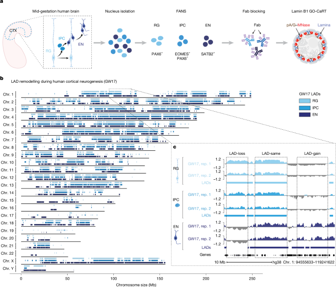
Snap-frozen cortical tissue was cut into small pieces and transferred to a prechilled 7 ml Dounce Tissue Grinder (Wheaton) containing 5 ml nucleus extraction buffer (NEB: 10 mM HEPES pH 7.4, 25 mM KCl, 5 mM MgCl2, 0.25 M sucrose, 0.1% Triton X-100, 1× Halt protease inhibitor cocktail (Thermo Fisher Scientific)). RiboLock (Thermo Fisher Scientific) was added to all the buffers to preserve RNA integrity. While still on ice, the tissue was dissociated with 5–6 strokes with loose pestle A followed by 8–10 strokes with tight pestle B, until no tissue pieces were visible. The sample was incubated on ice for 5 min after which it was transferred to a prechilled 15-ml conical tube. The sample was centrifuged at 500g for 10 min at 4 °C. The supernatant was removed, and the nuclear pellet was resuspended in 10 ml NEB without Triton X-100. The homogenate was passed through a 40 μm strainer into a 50 ml conical tube. Formaldehyde was added to a final concentration of 0.1% and the sample was incubated for 2 min at room temperature with rotation. Glycine was added to a final concentration of 75 mM to quench the reaction. BSA was added to a final concentration of 1% and the sample was centrifuged at 500g for 10 min at 4 °C. The supernatant was discarded, and the nuclear pellet was resuspended in 1 ml staining buffer (PBS containing 1% BSA). The nuclei were filtered through a 40 µm strainer into a 1.5 ml low-bind microcentrifuge tube and counted under a microscope. The nuclei were pelleted at 500g for 10 min at 4 °C and resuspended in 100–150 μl of staining buffer. Antibody staining was carried out for 1 h at 4 °C with rotation using the following antibodies: PAX6-Alexa Fluor 488 (BD Biosciences, 1:20), EOMES-PE-Cy7 (Invitrogen, 1:20) and SATB2-Alexa Fluor 647 (Abcam, 1:100). After staining, 900 μl of staining buffer was added and the samples were centrifuged at 500g for 10 min at 4 °C. The nuclear pellet was resuspended in 1 ml of staining buffer and centrifuged at 500g for 10 min at 4 °C. Nuclei were resuspended in 1–2 ml of staining buffer (depending on the starting material and yield) and filtered into a 70 µm mesh FACS tube (BD). DAPI was added at 1 μg ml−1 just before FANS. AbC Total Compensation capture beads (Thermo Fisher Scientific) were used for generating single-colour compensation controls. FANS was conducted on BD FACS Aria II Cytometer using a 70 μm nozzle. Sorted nuclei were collected in 5 ml tubes containing 300–500 μl of collection buffer (PBS containing 5% BSA and RNasin Plus RNase inhibitor (Promega)). Sorted nuclei were collected by centrifuging at 500g for 10 min at 4 °C and processed for downstream analyses (RNA-seq, Fab blocking and GO-CaRT/CUT&RUN).
Development of Fab blocking for antibody-based chromatin profiling
HEK293T cells were obtained from ATCC and authenticated at source. Cells were collected by trypsinization. Cells were fixed in 0.1% formaldehyde for 2 min at room temperature, followed by quenching in 0.1 M glycine (prepared in wash buffer). Cells were resuspended in 1 ml wash buffer (20 mM HEPES-KOH pH 7.5, 150 mM NaCl, 0.1% BSA, 0.5 mM spermidine and 1× Halt protease inhibitor cocktail) and centrifuged at 300g for 5 min. This step was repeated for a total of two washes. Cells were resuspended in wash buffer and aliquoted at 100 μl per tube into 0.5 ml PCR tubes (around 100,000 cells per experimental condition). Next, 8 μl of BioMagPlus concanavalin A beads (Polysciences) activated in binding buffer (20 mM HEPES-KOH pH 7.9, 10 mM KCl, 1 mM CaCl2 and 1 mM MnCl2) were added to each sample and rotated on a nutator for 10 min at room temperature. The cells were placed onto a magnet to clear and the liquid was removed. Cells bound to beads were resuspended in 50 μl cell permeabilization buffer (20 mM HEPES pH 7.5, 0.1 mM CaCl2, 3 mM MgCl2, 100 mM KCl, 0.05% digitonin (Sigma-Aldrich) and 1× Halt protease inhibitor cocktail) and incubated for 30 min at room temperature on a nutator. The supernatant was removed, and the beads were resuspended in 50 μl of antibody binding buffer (wash buffer containing 2 mM EDTA and 0.025% digitonin) containing an antibody against H3K27me3 (Cell Signaling Technologies, 1:100). IgG (Cell Signaling Technologies, 1:100) was included in parallel as a negative control. The samples were incubated overnight at 4 °C on a nutator. The next day, the supernatant was removed, and the beads were washed twice with 200 μl wash buffer containing 0.025% digitonin (Wash-Dig). The beads were resuspended in 50 μl of Wash-Dig containing monovalent anti-rabbit Fab fragments (Jackson Immuno) and incubated for 30 min at 4 °C. We tested the following Fab dilutions: 1:20, 1:50, 1:100, 1:250, 1:500, 1:1,000, 1:10,000, 1:50,000 and 1:100,000. We also tested three incubation times: 5 min, 15 min and 30 min. Dilutions of 1:250 and lower and incubation times of 5 min and below did not result in efficient blocking by the Fab fragments. After two washes with WaB-Dig (wash buffer containing 0.025% digitonin), the beads were resuspended in 50 μl of WaB-Dig containing pA/G-MNase (purified from Addgene plasmid 123461 at Macro Lab UC Berkley) and nutated at 4 °C for 1 h. The supernatant was removed, and the beads were washed twice with WaB-Dig. The beads were resuspended in 100 μl of WaB-Dig containing 2 mM CaCl2 to activate pA/G-MNase. The digestion was carried out for 30 min in a chilled metal block on ice. Digestion was stopped by adding 100 μl of 2× STOP (200 mM NaCl, 20 mM EDTA, 4 mM EGTA, 50 μg ml−1 RNase A, 40 μg ml−1 glycogen and 10 pg ml−1 heterologous DNA). The samples were incubated at 37 °C for 30 min to release pA/G-MNase-cleaved fragments. The contents of the tube were transferred to 1.5 ml Eppendorf tubes containing 2 μl each of 10% SDS and proteinase K (20 mg ml−1). The samples were incubated at 55 °C for 1 h to reverse cross-linking. DNA was extracted using the phenol–chloroform method. Purified DNA fragments were analysed by TapeStation High Sensitivity D1000 assay (Agilent).
To demonstrate that Fab blocking allows subsequent binding of another antibody for GO-CaRT/CUT&RUN analyses, we incubated the Fab-blocked H3K27me3 samples with an antibody against lamin B1. In brief, after Fab blocking of H3K27me3, the beads were resuspended in 50 μl of WaB-Dig containing lamin B1 antibody (Abcam, 1:100). The sample was nutated at 4 °C for 2 h. After two washes with WaB-Dig, the steps of pA/G-MNase binding, digestion and DNA extraction were carried out as described above. Purified DNA fragments were analysed by TapeStation High Sensitivity D1000 (Agilent).
Fab blocking followed by GO-CaRT and CUT&RUN analyses
Nuclei sorted by FANS were resuspended in PBS containing 1% BSA and divided into 100 μl aliquots in 0.5 ml PCR tubes. Next, 8 μl of activated BioMagPlus concanavalin A beads were added to each sample and rotated for 10 min at room temperature. The samples were placed onto a magnetic stand to remove the liquid. To block antibodies used in FANS, nuclei bound to concanavalin A beads were resuspended in 50 μl wash buffer (20 mM HEPES-KOH pH 7.5, 150 mM NaCl, 0.1% BSA, 0.5 mM spermidine and 1× Halt protease inhibitor cocktail) containing anti-rabbit and anti-mouse monovalent Fab fragments (Jackson Immuno) at 1:30. The samples were nutated at 4 °C for 30 min. The samples were then placed onto a magnetic stand to remove the liquid. The beads were resuspended in 200 μl wash buffer to remove excess Fab fragments. The beads were resuspended in 50 μl antibody binding buffer (wash buffer containing 2 mM EDTA) containing primary antibody of interest for GO-CaRT/CUT&RUN. Antibody incubation was carried out overnight on a nutator at 4 °C. The next day, the samples were briefly spun and placed onto a magnetic stand to remove the liquid. The samples were washed twice with 200 μl wash buffer. The beads were resuspended in 50 μl wash buffer containing pA/G-MNase (3 µg ml−1) and rotated for 1 h at 4 °C. The samples were briefly spun and placed onto a magnetic stand to remove the liquid. The samples were washed twice with 200 μl wash buffer. The beads were gently resuspended in a 100 μl wash buffer containing 2 mM CaCl2 (to activate pA/G-MNase) and placed in a prechilled metal block on ice. Digestion was carried out for 30 min and stopped by adding 100 μl of 2× STOP (200 mM NaCl, 20 mM EDTA, 4 mM EGTA, 50 μg ml−1 RNase A, 40 μg ml−1 glycogen and 10 pg ml−1 heterologous DNA). The samples were then incubated at 37 °C for 20 min to release pA/G-MNase cleaved fragments. Next, 2 μl SDS (10%) and 2 μl proteinase K (20 mg ml−1) were added to each sample and the sample was incubated at 55 °C for 1 h. DNA was extracted using the phenol–chloroform method. Purified DNA fragments were analysed using the TapeStation High Sensitivity D1000 assay (Agilent).
Lamin B1 GO-CaRT–seqChIP
iPS-cell-derived NPCs were collected with Accutase and the cell pellet was resuspended in 1 ml ice-cold PBS. The cells were centrifuged at 500g for 3 min at 4 °C and the cell pellet was resuspended in 1 ml of nucleus-isolation buffer (NIB: 10 mM HEPES-KOH pH 7.9, 10 mM KCl, 0.1% NP-40, 0.5 mM spermidine and 1× Halt protease inhibitor cocktail) and incubated for 10 min on ice. The nuclear pellet was collected by centrifuging at 600g for 3 min at 4 °C and again resuspended in 1 ml of NIB to wash. The nuclear pellet was collected by centrifugation at 600g for 3 min and resuspended in 100 μl of NIB.
Lamin B1 GO-CaRT was performed as described above, and lamin-B1-enriched chromatin released into the soluble fraction was used for subsequent sequential (two-step) ChIP. Lamin B1 GO-CaRT was performed in twelve 0.5 ml PCR tubes each containing around 500,000 nuclei (total of around 6 million nuclei) in 100 μl of NIB each. For the antibody and pA/G-MNase binding steps, the volumes were scaled up from 50 μl to 100 μl. The reaction was stopped using 2× STOP containing lamin B1 blocking peptide (Abcam) at 15 μg ml−1 followed by incubation at 37 °C for 15 min. The samples from 12 PCR tubes were pooled together and centrifuged at 14,000g for 5 min at 4 °C. The supernatant containing lamin-B1-associated DNA fragments was divided into 300 μl aliquots for the first ChIP. One aliquot was kept as input and stored at −20 °C for later use. For each ChIP, we used lamin B1 GO-CaRT supernatant from around 750,000 nuclei. The samples were incubated with antibodies against lamin B1 (1:100), H3K4me3 (1:100), H3K27me3 (1:50) and IgG control (1:100) at 4 °C overnight with rotation. The next day, protein A Dynabeads (Thermo Fisher Scientific) were equilibrated by washing twice in 1 ml wash buffer containing 0.05% Tween-20 (wash + Tween). The beads were resuspended in wash + Tween in the original volume of beads. Then, 15 μl of equilibrated beads was added to each ChIP reaction and the samples were incubated at 4 °C for 1 h. The samples were washed twice with 1 ml wash + Tween by rotating for 5 min at room temperature. The tubes were placed on the magnet to remove the supernatant. At this point, the controls for the first ChIP (lamin B1 GO-CaRT–lamin B1 ChIP and lamin B1 GO-CaRT–IgG ChIP) have finished processing and these samples were resuspended in 200 μl of wash + Tween and stored at −20 °C for DNA extraction later. For H3K4me3 and H3K27me3 first ChIP samples, chromatin was eluted by resuspending the beads in 150 μl of wash + Tween containing the corresponding blocking peptides (10 μg ml−1) for H3K4me3 (EpigenTek) and H3K27me3 (EpigenTek), respectively. The samples were incubated at 4 °C for 2 h. For H3K4me3 and H3K27me3, the first ChIP was carried out in two tubes so the total volume of eluted chromatin was 300 μl. The samples were placed on the magnet and the supernatant containing eluted chromatin from the first ChIP was transferred to a new 1.5 ml tube. A 100 μl aliquot of eluted chromatin was saved as the first ChIP input. To the remaining 200 μl of chromatin, an antibody of interest for the second ChIP (H3K27me3 if the first ChIP was done with H3K4me3 or H3K4me3 if the first ChIP was done with H3K27me3) was added and the samples were rotated at 4 °C overnight. The next day, 15 μl of equilibrated protein A Dynabeads were added to each second ChIP sample and incubated at 4 °C for 1 h. The samples were washed twice with 1 ml wash + Tween by rotating for 5 min at room temperature. The tubes were placed on the magnet to remove the supernatant and the beads were resuspended in 200 μl of wash + Tween. To each sample (including samples from first ChIP and inputs), 2 μl of 10% SDS and 2 μl of 20 mg ml−1 proteinase K were added. The samples were vortexed and incubated at 55 °C for 1 h. DNA was extracted using the phenol–chloroform method.
Library preparation and sequencing
Sequencing libraries for GO-CaRT, CUT&RUN and lamin B1-GO-CaRT–ChIP-reChIP were generated using KAPA HyperPrep Kit (Roche) according to the manufacturer’s instructions. The libraries were amplified for 12–14 PCR cycles. DNA fragments were quantified by Tapestation D1000 (Agilent) and Qubit high sensitivity dsDNA (Invitrogen) assays, pooled and sequenced by 150-bp paired-end sequencing on the NovaSeq 6000 or NovaSeq X Plus systems (Illumina).
CUT&Tag
CUT&Tag in iPS-cell-derived NPCs and neurons was performed as previously described, with modifications43. In brief, iPS-cell-derived NPCs and neurons were collected by scraping in 1× NIB, triturated and incubated on ice for 10 min. Cells were then centrifuged at 500g for 5 min at 4 °C. Nuclei were resuspended in PBS before fixation with 0.1% formaldehyde for 2 min. Fixation was quenched with 75 mM glycine for 2 min on ice. Nuclei were centrifuged and resuspended in wash buffer 1 (20 mM HEPES pH 7.4, 150 mM NaCl, 0.5 mM spermidine, 1× Halt protease inhibitor cocktail). For each reaction, 100,000 cells were counted for binding to activated concanavalin A magnetic beads and incubated with primary antibodies overnight on a nutator at 4 °C. On day 2, the samples were placed onto a magnetic stand and washed three times with wash buffer 1. The beads were resuspended in 50 µl wash buffer 1 containing secondary antibodies at a 1:100 dilution and nutated for 1 h at 4 °C. After clearing on a magnetic stand, the beads were washed twice with wash buffer 1, resuspended in 50 µl wash buffer 2 (20 mM HEPES pH 7.4, 300 mM NaCl, 0.5 mM spermidine, 1× Halt protease inhibitor cocktail) containing protein-A-fused preloaded Tn5 transposase at 1:100 dilution, and nutated for 1 h at 4 °C. The samples were then washed twice with wash buffer 2 and resuspended in 50 µl wash buffer 1 containing 10 mM MgCl2 for tagmentation at 37 °C for 1 h. The tagmentation reaction was quenched by addition of 1.7 μl 0.5 M EDTA, 0.5 μl 10% SDS and 0.5 μl 20 mg ml−1 proteinase K for 1 h at 55 °C. DNA was phenol–chloroform extracted and resuspended in 1× TE buffer (pH 8.0). DNA was PCR amplified by adding 2 µl each of 10 µM universal i5 primer and uniquely barcoded i7 primer44, along with 25 µl NEBNext High-Fidelity 2× PCR Master Mix (NEB). PCR products were size-selected with SPRIselect beads (Beckman Coulter) to recover DNA fragments over 75 bp according to the manufacturer’s instructions. Eluted DNA fragments were quantified using the Tapestation D1000 (Agilent) and Qubit high sensitivity dsDNA (Invitrogen) assays, pooled and sequenced by 150-bp paired-end sequencing on the NovaSeq 6000 system (Illumina).
GO-CaRT, CUT&RUN and CUT&Tag data processing and domain calling
Sequencing reads were first trimmed to remove adapters and low-quality sequences using Trim Galore (v.0.4.5, https://bioinformatics.babraham.ac.uk/projects/trim_galore) with the default parameters. Trimmed reads were mapped to the human reference genome (hg38) using Bowtie2 (v.2.3.5.1). PCR duplicates were removed using Picard tools (v.1.141, https://broadinstitute.github.io/picard). Post-alignment reads with a mapping quality score of less than 30 were filtered out using SAMtools (v.1.12). For lamin B1/SON, filtered .bam files were downsampled to match IgG read depth. For visualization and further analysis, downsampled .bam files of lamin B1/SON were converted to log2-normalized bigwig files using the deepTools (v.3.5.5) bamCompare function with the default parameters with IgG as the control. For visualization of histone modifications, bigwig files were generated using deepTools bamCoverage with the default parameters. Subsequent analysis and visualization of epigenomic data was performed in deepTools, bedtools (v.2.31.0) and ggplot2 (v.4.0.2).
Domain and peak calling
LADs were called by a two-state hidden Markov model in pomegranate (v.1.1.2) as previously described45. The model was called on the median of two biological replicate IgG-normalized bigwig files using a 30 kb bin size. For SPAD calling, bigwig files were converted to bedgraph format. The bedtools map function was then used to calculate average scores across 10 kb bins. Domains were called on the resulting bedgraph using SEACR (v.1.3) in the ‘non stringent’ mode by selecting the top 25% of regions by area under the curve. To ensure robustness of SPAD calls, only regions present in both biological replicates were used for further analysis. LADs/SPADs overlapping with blacklisted regions46 were discarded.
H3K4me3 and H3K27me3 peaks were called on the median of two biological replicates using MACS2 (v.2.1.2) with IgG as a control with options ‘–broad –q 0.05’ in addition to the default parameters. Identified peaks were annotated on the basis of the transcription start site (TSS) using Homer (v.4.11). All intersection calculations were performed using bedtools. Promoters were defined as ±1 kb around the TSS of the longest transcript of each gene. Bivalent promoters were those that contained peaks for both H3K4me3 and H3K27me3.
RNA-seq
Total RNA was extracted from RG, IPC and EN nuclei using the Quick-RNA FFPE RNA extraction kit (Zymo). Total RNA from NPCs and induced neurons was extracted using the Direct-Zol RNA Miniprep kit (Zymo). The samples were depleted of ribosomal RNA (rRNA) using the NEBNext rRNA Depletion kit v2 (NEB). Libraries for sequencing were prepared using the NEBNext Ultra II directional library kit (NEB) according to the manufacturer’s protocol. Libraries were quantified by TapeStation D1000 (Agilent) and Qubit high sensitivity dsDNA (Invitrogen) assays, pooled and sequenced on the NovaSeq 6000 or NovaSeq X Plus systems (Illumina).
Gene expression analyses
All fastq files were aligned to the reference genome (GENCODE GRCh38) by STAR v.2.7.11b after adaptor trimming. Reads that passed quality control and with read length greater than 50 bp were carried over for downstream counting after mapping to a gene transfer format (GTF) file. Read counts were calculated using featureCounts (v.2.24.0). Transcripts per million values were calculated using RSEM to estimate the absolute expression level for each gene. For heat-map plotting, raw read counts were normalized using DESeq2 (v.1.36.0) to account for sequencing depth and RNA composition between samples. RNA-seq data obtained in this study from GW20 RGs, IPCs and ENs, and GW17 RGs, IPCs and ENs from published datasets47, were integrated together into the analysis. The number of annotated transcripts for each gene in LAD, non-LAD/non-SPAD and SPAD was extracted from the Ensembl BioMart database using the biomaRt R package.
GO analyses
For a given list of genes of interest, GO analysis was performed using clusterProfiler (v.4.4.4) using the default parameters. GO terms with false-discovery rate < 0.05 were designated as significant functional enrichment. GO enrichment analyses was performed using over-representation analysis, corresponding to the one-sided Fisher’s exact test, to identify over-represented functional categories. P values were adjusted using the Benjamini–Hochberg procedure. Statistical significance was defined as an adjusted P < 0.05 and q < 0.05.
Statistics and data reproducibility
All GO-CaRT and RNA-seq experiments in the human cortex were performed using at least two biological replicates per gestational age. No statistical test was used to predetermine sample size. P values were derived from Wilcoxon’s rank-sum tests for genomic and transcriptomic comparisons throughout the study.
Integration of published GRO-seq data
iPS-cell-derived NPC fastGRO data from a previous study31 were obtained from GEO (GSE143844). Raw reads were processed using cutadapt (v.4.4) to remove adapters, low-quality bases and short reads. Cleaned reads were aligned to the hg38 or dm3 reference genome using STAR (v.2.7.11b) in 2-pass mode. Alignment files were normalized to the number of reads uniquely mapped to the Drosophila dm3 spike-in genome and converted to strand-specific bigwig files using deepTools (v.3.5.5). For downstream analysis, genes with a total length shorter than 400 bp were excluded. To quantify transcriptional activity, GRO-seq read densities at the TSS (±150 bp) and the gene body (TSS + 300 bp to the transcription end site) were calculated using bedtools (v.2.31.0) and visualized with ggplot2.
DNA-FISH combined with immunocytochemistry and image analyses
DNA-FISH with immunocytochemistry for lamin B1 and SON was performed as described previously8 with slight modifications. In brief, the medium was removed from the chambered coverglass containing dissociated cells from germinal zone and cortical plate and cells were fixed with Histochoice MB (Sigma-Aldrich) for 30 min at room temperature. Cells were washed three times for 10 min each with D-PBS. Cells were permeabilized and blocked in D-PBS containing 5% NGS and 0.5% Triton X-100 for 1 h at room temperature. Cells were incubated in antibody binding solution (5% NGS and 0.1% Triton X-100 in D-PBS) containing lamin B1 or SON antibodies at a 1:500 dilution. The next day, cells were washed three times with D-PBS and incubated with Alexa Fluor 647 anti-rabbit secondary antibody (1:500) for 2 h at room temperature. After three washes with D-PBS, cells were post-fixed in Histochoice MB for 20 min at room temperature. Cells were washed twice with D-PBS, and 0.7% Triton X-100 was included in the third wash to repermeabilize the cells. After a quick wash with D-PBS, cells were treated with an ethanol gradient: 70%, 85% and 100% ethanol each for 2 min at room temperature. The chambered coverglass was allowed to dry at 45 °C until all ethanol evaporated. A humidified chamber was prepared using 50% formamide/2× SSC solution in a dark box and preheated to 83 °C. The DNA-FISH probe (Empire Genomics) mixture (3 µl probe, 17 µl hybridization buffer, 5 µl denaturation buffer (70% formamide, 2× SSC pH 7–8)) was added to the cells and the chambered coverglass was placed in the pre-heated humidified chamber and denatured at 83 °C for 10 min. The samples were transferred to a 37 °C oven and incubated for 16–24 h. The next day, wash solution 1 (0.3% NP-40 and 0.4× SSC) prewarmed to 73 °C was added and incubated for 3 min at room temperature. A second wash was performed with wash solution 2 (0.1% NP40 and 2× SSC) for 3 min at room temperature and the coverglass was allowed to dry in the dark. Cells were stained with DAPI (2 µg ml−1) for 10 min at room temperature, washed with D-PBS and water and mounted with ProLong Glass Antifade Mountant (Invitrogen). The samples were left in the dark overnight at room temperature to cure mountant before imaging.
Imaging was performed using the Leica TCS SP5X confocal microscope with a ×63 oil immersion objective, with a z-stack collected for each channel (step size 0.2 μm). Images were acquired with LAS X software (v.1.9.0, Leica). ImageJ software (v.2.0.0-rc-65/1.52q, build: 961c5f1b7f) was used to process images. Nuclear lamina and speckles were identified by lamin B1 and SON immunostaining, respectively. 3D reconstructions of cells were conducted in Imaris x64 (v.9.2.1) software (Bitplane). DNA-FISH dots for probe signals were created at the location of the highest fluorescence intensity using the Spots tool. The spot diameter ranged from 350 nm to 400 nm. Nuclear lamina/speckle surfaces were automatically detected from lamin B1/SON immunostaining using the Surfaces tool. The distance from the centre of the FISH spot to the closest lamina/speckle surface was quantified using the Measurement Points tool. For each experimental condition, 40–50 nuclei were analysed. In the case that the generated DNA-FISH spot was embedded in the laminar/speckle surface, the distance to the lamina/speckle was quantified as zero.
Plasmid construction
LacI plasmids were constructed using the In-Fusion Snap Assembly method (Takara). GFP-LacI-NLS transgene (derived from Addgene plasmid 183920, from K. Rippe) was inserted into the pCMV-N-Flag vector to generate pCMV-Flag-GFP-LacI. Human lamin A CDS (derived from Addgene plasmid 129275, from A. Ting) was inserted C terminally to LacI to generate pCMV-Flag-GFP-LacI-LMNA. pEF-1α-pLacO-RFP was purchased from Addgene (179507, from Y. Hiraoka).
HEK293T cell culture, plasmid biotinylation and transfection
HEK293T cells (ATCC) were cultured in DMEM supplemented with 10% FBS and 1% antibiotic-antimycotic (Sigma-Aldrich). pEF-1α-pLacO-RFP plasmid was biotinylated and repurified using the Label-IT Tracker Intracellular Nucleic Acid Localization Kit, Biotin (Mirus) according to the manufacturer’s instructions. Biotinylated and unmodified plasmids were transfected into HEK293T cells using Lipofectamine 3000 Transfection Reagent (Invitrogen) and collected 3 days after transfection. For drug treatment experiments, cells were treated with 5 µM EPZ-6438 or an equivalent volume of DMSO for 4 days before transfection. The drug was replenished every other day and treatment was continued after transfection until collection.
Immunofluorescence and plasmid visualization
HEK293T cells transfected with GFP-LacI/GFP-LacI-LMNA and biotinylated pEF-1α-pLacO-RFP were fixed with 4% paraformaldehyde for 20 min at room temperature. Cells were washed in PBS and free aldehydes were quenched with 125 mM glycine in TBS. Cells were then permeabilized and blocked (5% normal donkey serum, 5% normal goat serum (Jackson Immunoresearch), 0.25% Triton X-100 in PBS) for 1 h at room temperature. Cells were incubated with rabbit anti-lamin B1 (Abcam, ab16048, 1:250) and chicken anti-GFP (Abcam, ab13970, 1:500) primary antibodies, diluted in antibody buffer (5% normal donkey serum, 5% normal goat serum, 0.1% Triton X-100 in PBS) overnight at 4 °C. The next day, cells were washed four times for 5 min in PBST followed by incubation in antibody buffer with Streptavidin Alexa Fluor 647, donkey anti-rabbit Alexa Fluor 555 and goat anti-chicken Alexa Fluor 488 (Invitrogen, all 1:500) for 2 h at room temperature in the dark. Cells were then washed five times in PBST, incubated with 1 µg ml−1 DAPI in PBS for 5 min, and mounted with ProLong Glass Antifade Mountant. The slides were imaged on the Leica Stellaris confocal microscope with a ×63 objective. The plasmid distance to the lamina was analysed in Imaris as described for DNA-FISH.
Western blotting
Cultured cells were washed with ice-cold PBS and collected on ice in RIPA buffer (50 mM Tris-HCl pH 7.5, 150 mM NaCl, 1 mM EDTA, 0.1% SDS, 0.5% sodium deoxycholate) supplemented with 1× Halt protease inhibitor cocktail. The lysates were sonicated on ice and centrifuged at 3,000g for 10 min at 4 °C. Then, β-mercaptoethanol was added to supernatants to a final concentration of 5%, and the samples were mixed with LDS sample buffer (Thermo Fisher Scientific) and boiled for 5 min. Next, 20 μg of protein was resolved by SDS–PAGE. Protein was transferred onto PVDF membranes, which were blocked in 5% BSA in TBS with 0.1% Tween-20 (TBST) followed by overnight incubation in primary antibodies (diluted 1:1,000 in blocking buffer) at 4 °C. The membranes were washed in TBST and incubated in HRP-tagged anti-rabbit secondary antibody (Cell Signaling, diluted 1:5,000 in blocking buffer) for 1 h at room temperature. Proteins were visualized using SuperSignal West Pico Plus Chemiluminescent Substrate (Thermo Fisher Scientific) and imaged on the LICOR Odyssey XF system.
RNA isolation and RT–qPCR
Cells were collected in TRIzol (Invitrogen) and RNA isolated using the Direct-zol RNA Miniprep kit (Zymo) according to the manufacturer’s instructions. cDNA was reverse transcribed from 0.5 µg RNA using the Transcriptor First Strand DNA Synthesis Kit (Roche) with oligo-dT primers according to the manufacturer’s instructions. qPCR with reverse transcription (RT-qPCR) was performed in technical triplicate on the LightCycler 480 Instrument (Roche) using the LightCycler 480 SYBR Green I Master Mix (Roche) according to the manufacturer’s instructions. Triplicate values were averaged and RNA expression was determined using the ΔΔCt method using GAPDH as the loading control.
Chromatin immunoprecipitation
HEK293T cells in 10 cm dishes were fixed with 1% formaldehyde for 10 min at room temperature, quenched for 5 min with 125 mM glycine and washed twice in PBS. Cells were collected in lysis buffer 1 (50 mM HEPES pH 7.5, 140 mM NaCl, 1 mM EDTA, 10% glycerol, 0.5% NP-40, 0.25% Triton X-100 and 1× Halt protease inhibitor cocktail (Thermo Fisher Scientific, included in all buffers)) and freeze–thawed in liquid nitrogen. During thawing, protein G Dynabeads (Invitrogen) were washed and incubated with 1 µg normal rabbit IgG, rabbit anti-H3 or rabbit anti-H3K27me3 (Cell Signaling) in 0.5% BSA/PBS for 2–4 h at 4 °C. Thawed cells were Dounce homogenized 20 times with type B pestle to liberate nuclei, which were pelleted at 500g for 5 min. The nuclear pellet was resuspended in lysis buffer 2 (10 mM Tris-HCl pH 8, 200 mM NaCl, 1 mM EDTA, 0.5 mM EGTA), rotated at 4 °C for 10 min and centrifuged at 500g for 5 min. The washed pellet was resuspended in lysis buffer 3 (10 mM Tris-HCl, pH 8, 100 mM NaCl, 1 mM EDTA, 0.5 mM EGTA, 0.1% sodium deoxycholate) and sonicated in a Bioruptor ice bath sonicator on high power for 25 cycles (30 s on/30 s off). Before the final ten cycles, Triton X-100 was added to a final concentration of 1%. Sonicated lysates were cleared at 16,000g for 10 min at 4 °C. The supernatant was split equally among three tubes (10% split sample volume saved as input), mixed with appropriate bead–antibody complex and rotated overnight at 4 °C. The next day, beads were washed five times for 2 min at room temperature in wash buffer 1 (50 mM HEPES pH 7.5, 500 mM LiCl, 1 mM EDTA, 1% NP-40, 0.7% sodium deoxycholate) and once in wash buffer 2 (10 mM Tris-HCl pH 8, 1 mM EDTA, 50 mM NaCl). Chromatin was eluted from beads by shaking at 65 °C for 30 min in elution buffer (50 mM Tris-HCl pH 8, 10 mM EDTA, 1% SDS) then transferred to a fresh tube. NaCl (final concentration 540 mM) and proteinase K (0.2 mg ml−1) were added to ChIP and input samples followed by overnight incubation at 65 °C. The next day, the samples were treated with RNase A (0.1 mg ml−1) for 30 min at 37 °C. DNA was then purified by 1.8 AMPure XP Bead (Beckman) cleanup and eluted in 0.1× TE buffer. qPCR was performed as described above.
Reporting summary
Further information on research design is available in the Nature Portfolio Reporting Summary linked to this article.

