


How a single fertilized cell gives rise to the intricate architecture of the human body is one of biology’s most enduring questions. Writing in Nature, Pan et al.1 present a high-resolution spatiotemporal atlas of whole human embryos, capturing the molecular details of early organ formation. With this resource, they provide a powerful framework for connecting genetic variation to human disease.
Read the paper: Spatiotemporal transcriptome atlas of human embryos after gastrulation
The most decisive events in human development occur early in pregnancy, during embryogenesis in the first trimester, when the initially simple embryo rapidly gains cellular and molecular complexity. During this brief window, the body plan is established and the process of organogenesis begins. This is the developmental stage at which the foundations of all major organ systems are laid out and when many congenital disorders originate.
Yet much of our understanding of these processes still comes from model organisms such as mice and zebrafish (Danio rerio), reflecting the practical and ethical challenges of studying human embryos. Even though single-cell genomics efforts and global initiatives such as the Human Developmental Cell Atlas2 have begun to catalogue cell types across individual embryonic organs, a comprehensive view of the entire human embryo during organogenesis — including the spatial coordination of developing tissues — has remained elusive2,3. This gap is particularly important because programs of gene regulation begin to diverge across vertebrates during this stage, limiting the extent to which model organisms can fully explain the origins of human developmental diseases4.
Developmental maps of the brain trace when cell types emerge
Pan and colleagues address this by mapping gene expression during human organogenesis with incredible spatial and cellular detail. By placing molecular programs in their anatomical context, their resource provides a framework for linking the regulation of genes during development to the origins of human disease.
Part of the challenge is that most existing data sets lack spatial information. Yet developmental programs unfold in space as well as time, and the positional context of gene expression is particularly important in anatomically diverse tissues such as muscle, bone and epithelia (the tissue that lines surfaces in the body). During the past decade, scientists have begun to address this limitation using a technology known as spatial transcriptomics, which enables gene expression in cells to be measured while preserving the location of those cells in their native tissue.
One such method is called Stereo-seq, which was developed by some of the authors of the current work. It uses a patterned chip to record the spatial origin of sequenced RNA transcripts with a resolution of about 25 micrometres — roughly the size of a human cell. This technology has led to the creation of spatial maps of organogenesis in mice5 and of brain regeneration in axolotls (Ambystoma mexicanum)6. Pan and colleagues now extend this approach to thin sections of developing human embryos, offering an unprecedented view of human organogenesis in its spatial context (Fig. 1).
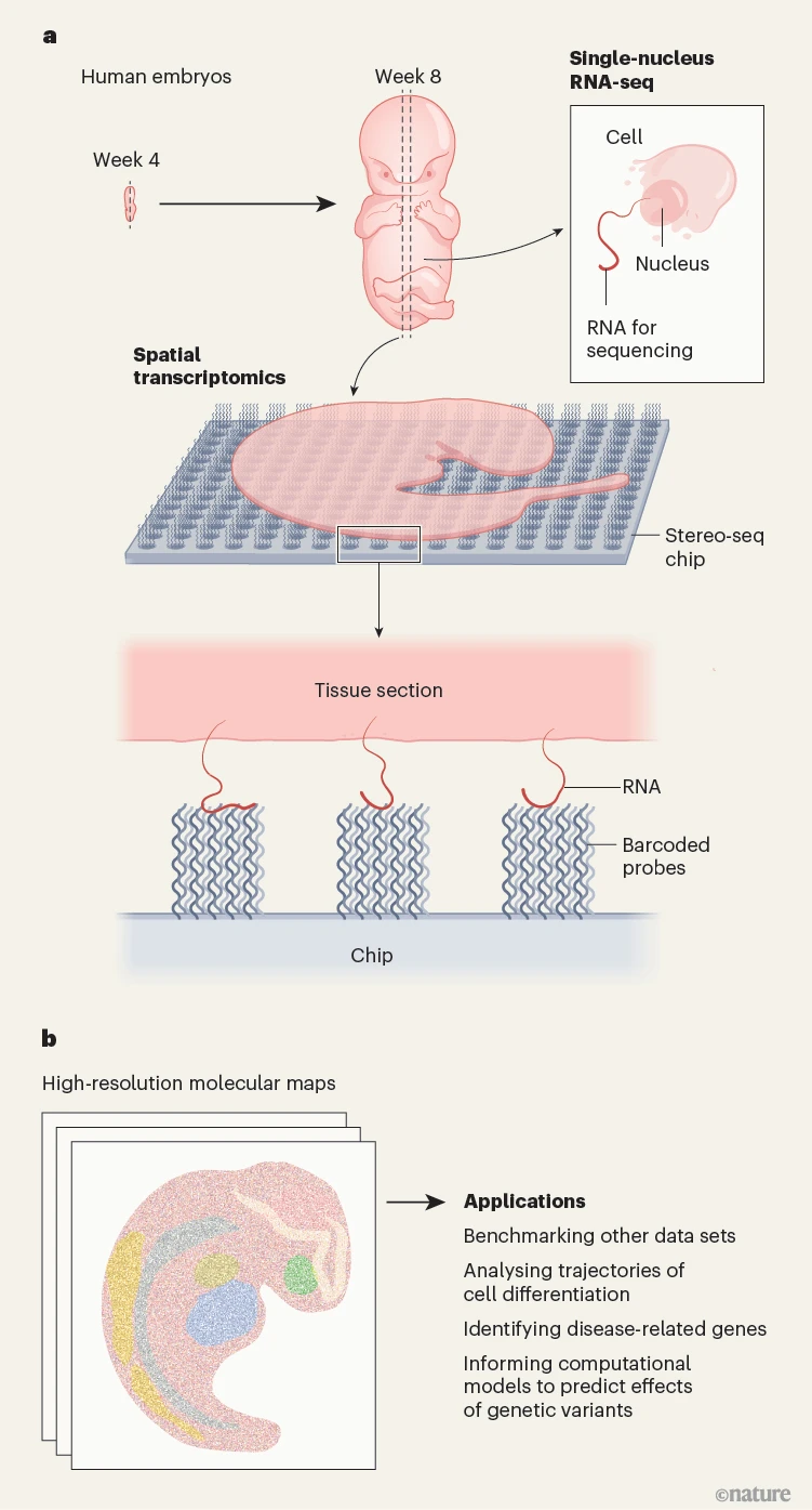
Figure 1 | Generating a gene-expression atlas of developing human embryos. a, Using donated human embryos spanning four to eight weeks post-conception, Pan et al.1 created spatial maps of gene expression that capture the molecular details of organ formation. They used a spatial transcriptomics technique called Stereo-seq, which involves laying thin sections of tissue on a patterned chip containing barcoded probes. The probes capture RNA transcripts from the tissue, and the barcodes enable the transcripts to be mapped back to their original position in the tissue after RNA sequencing. The authors integrated this information with single-nuclei RNA-sequencing data from the sections. b, The resulting high-resolution molecular maps are publicly available and have several potential applications relevant to human development and disease.
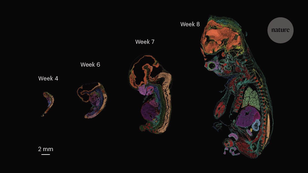
The authors analysed 13 whole human embryos, spanning four to eight weeks post-conception, that were donated after voluntary termination of pregnancy. With these valuable samples, they could chart the emergence of cell types and gene programs across 50 developing organs (Fig. 2). The combination of precise temporal staging and spatial transcriptomic resolution enabled the authors to capture key developmental transitions as they unfold in the intact embryo.
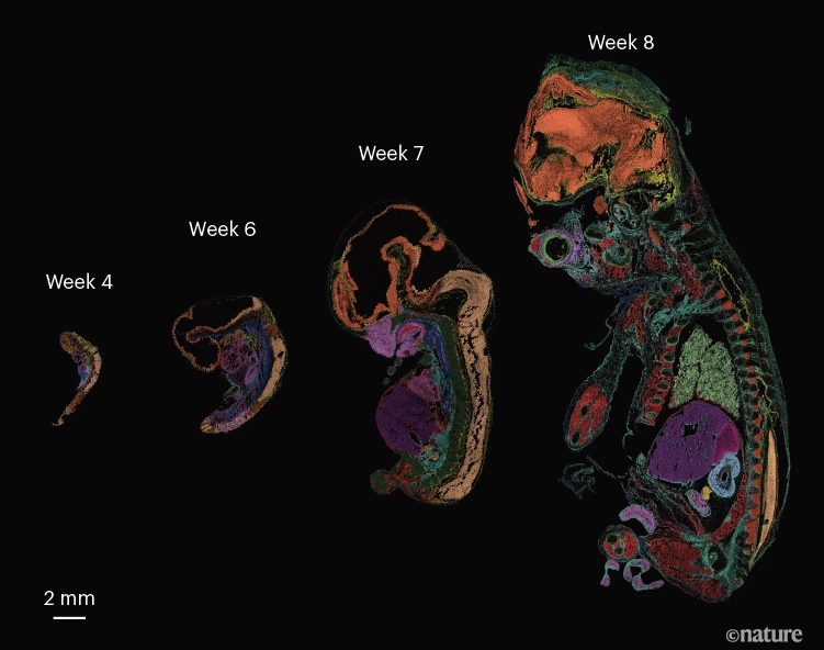
Figure 2 | Spatial transcriptomic maps of human embryos. Each spot is coloured according to the tissue type inferred from the gene-expression profile measured at that location. Each of the 50 colours represents a different developing organ. (Adapted from Fig. 1b of ref. 1.)
In the developing heart, for example, the atlas pinpoints the formation of the sinoatrial node — a small structure that contains specialized muscle cells and acts as the heart’s pacemaker — revealing transcriptional programs that underlie its specification and maturation. In the eye, the data uncover divergent gene-regulatory trajectories that guide immature cells called retinal progenitors towards distinct cell fates, including pigment-producing ‘melanocytes’ and various neuronal cells. More broadly, the study documents a continuous increase in cellular diversity as development progresses, accompanied by the emergence of increasingly specialized gene-expression programs for functions such as muscle contraction in skeletal muscle and visual perception in the retina. Together, these observations illustrate how spatially resolved molecular maps can illuminate the coordinated gene-regulatory programs that shape the human body.
But what can such an atlas reveal about human diseases? By identifying where and when genes are active during embryogenesis, spatial maps of development provide a powerful framework for interpreting genetic variants found in people with congenital conditions. Pan and colleagues demonstrate this by examining the expression of nearly 2,000 genes that are associated with developmental disorders. Many of these genes are enriched in the tissues that would be expected on the basis of the disorder’s clinical manifestations. For example, the genes CRYBA1 and BFSP2, which are mutated in congenital cataracts, are enriched in the developing eye. Importantly, the atlas also highlights cases in which human developmental programs diverge from those of model organisms. For instance, the liver-enzyme gene ARG1 is enriched at different times in humans and mice, cautioning against direct extrapolation from animal models.
Primate embryo model leaps across developmental boundaries
Beyond inherited disorders, the atlas also offers insights into infectious diseases. For example, infection with Zika virus during pregnancy can affect brain development in the fetus. The authors identified the expression of receptors for the virus in the brains and spinal cords of the embryos, which might explain the susceptibility of fetal brains to infection during gestation.
More broadly, such a data set provides a foundational reference for human biology, presenting a scaffold on which to map complementary types of data. The authors take this approach by projecting RNA-sequencing data from single nuclei onto the spatial transcriptomic maps to overcome the limited sensitivity of Stereo-seq. This approach could enable the detection of abnormal transcriptional signatures that are seen in diseased tissue or offer a benchmark to optimize emerging in vitro models of human embryos, known as embryoids. Finally, these richly annotated data sets provide an invaluable training ground for new computational models that aim to integrate different types of genomic data and predict the effects of genetic variation. In this way, developmental atlases such as this one could ultimately help to bridge the gap between genetic variation and human disease.
But as is often the case, there is always more to be learnt. Gene expression, measured by Stereo-seq, captures only one facet of development — the output of the genome rather than the regulatory mechanisms that drive it. Regulation of gene expression can be assessed by looking at the accessibility of chromatin, the packaged form of DNA, which reflects whether a region of the genome is participating in transcription7. Alternatively, it can be assessed by evaluating the epigenetic landscape (the pattern of chemical modifications) at non-protein-coding regions of the genome such as enhancer sequences. Integrating this kind of information will be essential for understanding how gene programs are established and disrupted in disease. Moreover, most spatial transcriptomic approaches capture tissues in 2D, missing aspects of cellular organization that occur in 3D. Advances in sequencing and imaging technologies, as well as their integration, promise to provide an increasingly complete view of human development.
For now, however, Pan and colleagues present an atlas that marks a milestone, opening a remarkable window into human organogenesis. Perhaps most importantly, they have made these long-awaited data available to the wider community of scientists and physicians who seek to understand the origins of human disease (see db.genomics.cn/hesta).
Competing Interests
The authors declare no competing interests.

