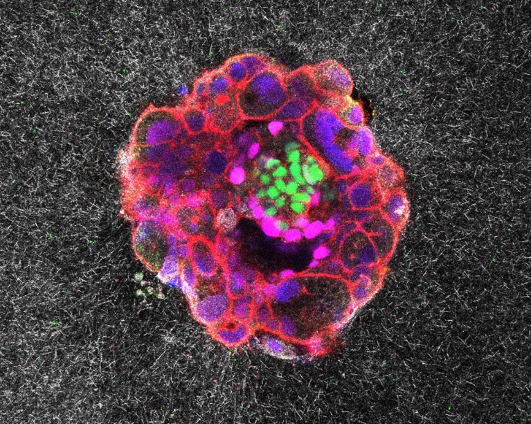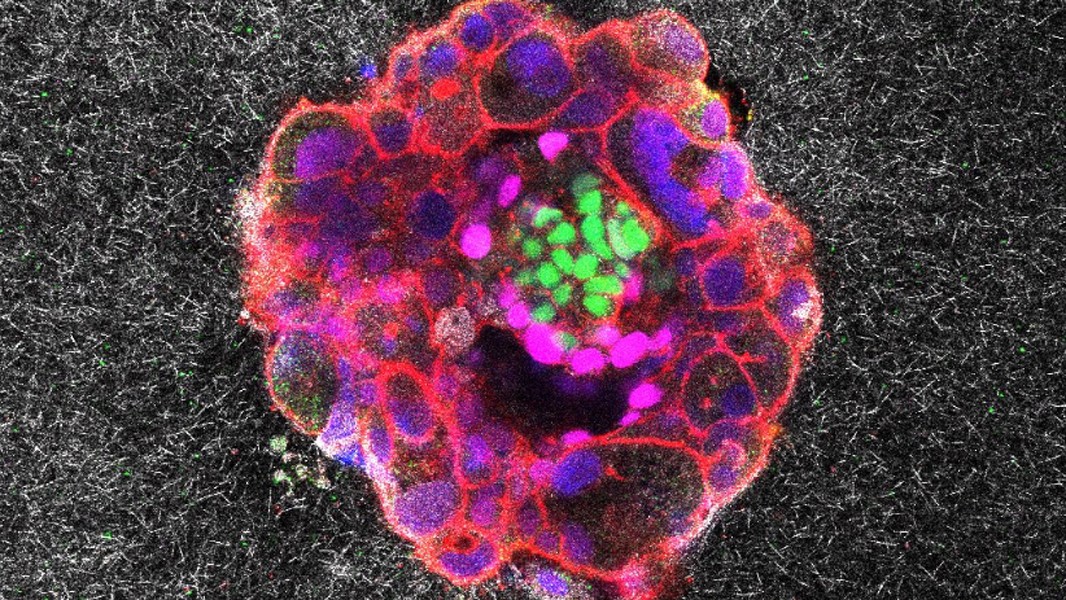
A microscopic view of a nine-day-old human embryo shows a protein found in embryonic stem cells in green, developing tissue in magenta and DNA in blue. Credit: Institute for Bioengineering of Catalonia (IBEC)
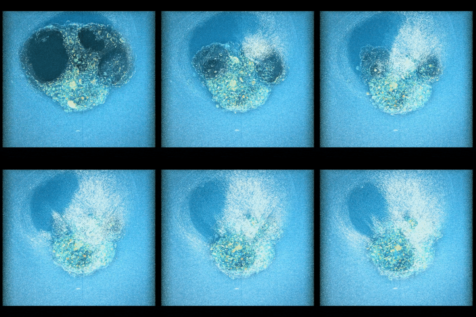
A time-lapse film offers a glimpse of a hidden milestone of human development: the moment when the newly formed embryo latches onto the uterine lining. Researchers have captured real-time footage of an embryo pulling on a high-fidelity replica of the lining to bury itself inside, effectively remodelling its new home.
The team reports its findings today in Science Advances1.
Hidden figures
The authors were inspired to simulate the implantation process because the actual events are so difficult to capture. “It’s very inaccessible because it’s all happening inside the mother,” says co-author Samuel Ojosnegros, a bioengineer at the Institute for Bioengineering of Catalonia (IBEC) in Barcelona, Spain. “It’s such an important process for human reproduction, but at the same time, we don’t have the technology to study it.”

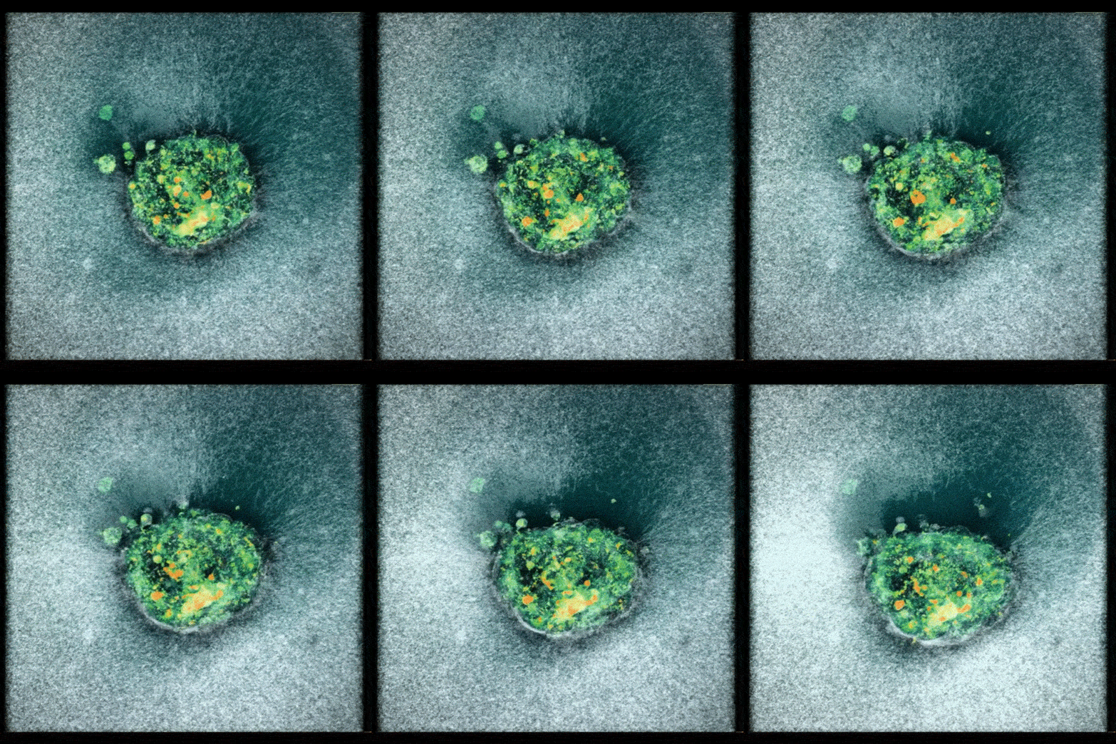
A human embryo contracts itself to minimize its exposure to the outside environment.Credit: Institute for Bioengineering of Catalonia (IBEC)
Although previous studies have investigated how human embryos interact with glass, the embryo can’t penetrate this material as it does real human tissue. So the team set out to create a more lifelike mock-up, devising a faux uterine lining from a gel rich in collagen and proteins that are crucial for embryonic development.
To shoot their stop-motion film, researchers placed human embryos donated by a local hospital near the gel. As the embryo attached to the ‘uterus’, the team used a microscope to capture an image about every 20 minutes for 16–24 hours, and then stitched the stills together.
Co-author Amélie Godeau, a biomechanics researcher at IBEC, was shocked to see how quickly the embryo burrowed down into the gel. “My first reflex was to think my experiment had gone wrong and there was some drift in the microscope,” Godeau says. By contrast, the team found that mouse embryos adhere to the surface of the uterus rather than embedding itself inside.

A human embryo plunges into a synthetic uterine lining tissue by applying force to pull the tissue apart.Credit: Institute for Bioengineering of Catalonia (IBEC)


