


A decade ago, Helena Jambor found herself struggling to understand the figures in the scientific papers she was reading during her postdoc. Jambor was studying how fluorescently labelled messenger RNA arranges itself inside embryos of the fruit fly Drosophila melanogaster and wanted to choose the images that would most clearly convey her findings. But she couldn’t find many helpful guidelines — or even good examples in the literature. “When you start out, you think you’re the one who’s stupid,” she says. “But after ten years in science, you realize, okay, now I’m an expert and I still don’t understand some of these figures.”
Jambor, now a biologist and visualization specialist at the Dresden University of Technology in Germany, realized that image presentation wasn’t just a problem in her own field. In 2021, when she and her colleagues analysed the figures of 580 biology papers in 15 top journals, they found that most contained poorly presented images (H. Jambor et al. PLoS Biol. 19, e3001161; 2021). Many panels lacked labels or scale bars, and annotation features, such as descriptive text and arrows, were often missing from the imaged objects. “I just think it’s sad if someone spends four years researching an amazing issue and publishes a paper and then has a figure that is not fully understandable,” she says. “It will inevitably reduce the readership.”
Will AI jeopardize science photography? There’s still time to create an ethical code of conduct
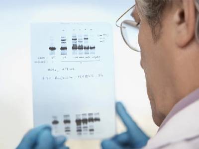
Whereas scientists receive extensive training on how to collect data, less work goes into teaching them how to showcase the information in publications, presentations and grant applications. For many types of figure panel, including photomicrographs and images of gel-based protein assays known as western blots, some photo editing is required to present them clearly. But there’s a fine line between clarifying and manipulating — as highlighted by increasing attention given to scientific misconduct in published papers.
Some journals and scientific societies are now developing guidelines to help researchers to present their hard-won data well while maintaining image integrity. The increased attention is a good thing, says Elisabeth Bik, a microbiologist and full-time image sleuth in San Francisco, California. “If you put the effort into those fronts, you can find [both errors and deliberate manipulations] before they get published.”
Slippery slope
Many aspects of editing are common sense, says Jana Christopher, an image-integrity analyst at the Federation of European Biochemical Societies, who is based in Heidelberg, Germany. For instance, it’s OK to increase the image’s contrast so that a cell or feature pops out against the background, but the background still has to be visible. Altering brightness or saturation is also acceptable as long as the modification is applied equally across the whole image. Cropping is fine if the process doesn’t remove elements that would change the interpretation of the image — after all, Christopher notes, selecting a particular field of view under the microscope is in itself a form of cropping and selective reporting. “The bottom line is that the images need to represent accurately what was observed experimentally,” she says.
Image annotation is essential. Jambor is shocked by how many photographs lack scale bars, for instance. “People seem to just think that it’s nice for decoration or something, but it’s actually essential,” she says, because biological scales vary widely. “If we blow the smallest cell up to the size of a ping-pong ball, the largest cell, in comparison, is the height of Mount Everest.”
How journals are fighting back against a wave of questionable images
Accessibility is also often overlooked. Jambor’s study found that some 20–50% of biological papers contain at least one figure that isn’t understandable for people who have red–green colour blindness, for instance. Showing coloured fluorescent images side by side instead of overlaid on one another — and annotating everything — helps to alleviate this problem. So does selecting a different colour palette or showing the images in greyscale. Popular image-processing software, such as ImageJ, includes tools that check whether images are suitable for people who are colour-blind.
But as helpful as such programs can be, the temptation to beautify images can be a slippery slope. Some researchers, for instance, will use editing software, such as Adobe Photoshop, to ‘clone’ parts of an image and paste them onto other sections to cover a crack or piece of dust in the sample. This calls the entire paper’s integrity into question, Bik says: “We’ll ask questions.”
As experimental technology changes, so do standards around the images they capture. “With any new technique, you have to come to an agreement on what is possible or ethical,” Bik says. For instance, 20 years ago it was considered acceptable to put pieces of different western blots next to one another and photograph them as one image. Nowadays, Bik says, this is frowned on and authors are expected to make it clear when this has been done. The cardinal rule is to show your work.
Colour me better: fixing figures for colour blindness
Some standards, however, remain constant, Bik notes. A 2004 paper in the Journal of Cell Biology — published just as tools such as Photoshop were becoming widely available to scientists — outlined guidelines that she says are still the gold standard for image analysis today (M. Rossner and K. M. Yamada J. Cell Biol. 166, 11–15; 2004). For instance, the study’s authors wrote that researchers shouldn’t selectively enhance specific features, for example by adding a black dot in Photoshop to make a particle more visible, or increase the number of pixels beyond those contained in the original image, effectively creating new data.
“Data must be reported directly, not through a filter based on what you think they ‘should’ illustrate to your audience,” the authors wrote, warning against the “tempting dangers of digital manipulation”. They added, “Just because the tools exist to clean up sloppy work digitally, that is no excuse to do sloppy work.”