Synthetic peptides
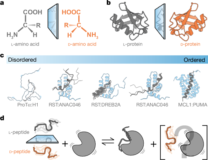
Synthetic l- and d-peptides of H1155â175, PUMA130â156, ANAC013254â274, ANAC046319â338 and DREB2A255â272 were purchased from Pepscan (now Biosynth) at a minimum purity of 95% and purified by HPLC. The d-peptides contain amino acid residues with a stereoisomeric d-form of each chiral carbon. The peptides were either resuspended in MilliQ H2O or in MilliQ H2O containing 50âmM NH4HCO3 and lyophilized repeatedly to remove leftover trifluoroacetic acid from the last purification step by the manufacturer. Peptides were then either resuspended directly in the buffer used for experiments or in H2O without 50âmM NH4HCO3 to measure the concentration. If no aromatic residue was present in the peptide sequence, the absorbance at 214ânm was used. The extinction coefficient was calculated using Bestsel51.
Expression and purification of proteins
15N-labelled and unlabelled full-length ProTα was expressed and purified as described22. The double-cysteine variant of ProTα (E56C/D110C) used in smFRET experiments was expressed and purified as described24, with some modifications. In brief, ProTα was dialysed against Tris buffer (50âmM Tris, 200âmM NaCl, 2âmM DTT, 1âmM EDTA; pH 8), during which the hexa-histidine tag was cleaved using HRV 3âC protease. Cleaved ProTα was purified further using Ni Sepharose Excel resin (Cytiva, formerly GE Healthcare) and a HiPrep Q FF column (Cytiva) with a gradient from 200âmM to 1âM NaCl. Buffer was exchanged (HiTrap Desalting column (Cytiva)) to labelling buffer potassium phosphate (100âmM, pH 7). 15N-labelled and unlabelled GSTâMCL1152â308 was expressed in BL21(DE3)pLysS Escherichia coli in the presence of ampicillin. Cells were grown at 37â°C in LB or M9 minimal medium (for 15N labelling) until OD600 reached 0.6, then induced with IPTG (1âmM final concentration) and collected after 4âh. The cell pellet was resuspended in Tris buffer (20âmM Tris, 100âmM NaCl; pH 8), then lysed by sonication. After pelleting again, the supernatant was applied to GST Sepharose beads (Cytiva), and GSTâMCL1152-308 was eluted using Tris-GSH buffer (20âmM Tris, 100âmM NaCl, 10âmM GSH; pH 8). The GST tag was removed using TEV protease (0.7âmg) overnight at room temperature. Final purity was reached using a Superdex 75 26/60 column (Cytiva), equilibrated with 50âmM phosphate buffer (pH 7). 13C,15N-labelled MCL1152â308 was expressed as described52 and purified as above. The expression and purification of 15N-labelled and unlabelled RCD1-RST499â572 were carried out as previously described28 with the lysis buffer changed to 20âmM Tris-HCl, pH 9.0, 20âmM NaCl. The buffer used in the last purification step by size exclusion chromatography on a Superdex 75 10/300 GL column (Cytiva) was the buffer described for the individual methods.
13C,15N-labelled ANAC046319â338 or ANAC013254â274 were expressed with a His6-SUMO fusion tag in BL21(DE3) E. coli in the presence of kanamycin (50âμgâmlâ1). Cells were grown in LB at 37â°C until OD600 reached 0.6 and the medium was changed to M9 minimal medium, followed by induction with IPTG to 1âmM final concentration and collected after incubation overnight at 16â°C. The cells were resuspended in lysate buffer (50âmM and 20âmM Tris-HCl for ANAC046319â338 and ANAC013254â274, respectively, pH 8.0, 300âmM NaCl) and sonicated. After the centrifugation, the lysate was purified using TALON resin equilibrated in the buffers just described. The fusion peptides were eluted with an equivalent buffer containing 250âmM imidazole. After a dialysis step into 20âmM Tris-HCl pH 8.0, 100âmM NaCl, the fusion tag was cleaved with ubiquitin-like-specific protease 1 (ULP1) (molar ratio between peptide and protease were 1:320 and 1:500 for ANAC046319â338 and ANAC013254â274, respectively) overnight at 4â°C. A second purification step with TALON resin was performed resulting in the peptides in the flowthrough. The purification of the peptides was finalized by size exclusion chromatography on a Superdex peptide 10/300 GL column (Cytiva) and freeze-dried to be resuspended in the desired buffer.
AlphaFold structure modelling
Protein interaction models of RCD1-RST499â572 in complex with ANAC046319â338 or ANAC013254â274 were generated using AlphaFold330 and analysed in PyMOL (The PyMOL Molecular Graphics System, version 3.0 Schrödinger, LLC.). The five generated models for each complex were assessed manually and compared with the secondary chemical shifts of Cα of the l-ligand recorded using ZZ-exchange or CEST (see NMR spectroscopy method). The structures agreeing with the experimental data were visualized in PyMOL or Chimera X53.
Far-UV CD spectropolarimetry
Far-UV CD spectra of l- and d-peptides of H1155â175, PUMA130â156, ANAC013254â274, ANAC046319â338, and DREB2A255â272 were measured on a Jasco 815 spectropolarimeter with a Jasco Peltier control in the range of 260â190ânm at 20â°C. Concentrations of peptides varied between 10â30âµM in either MilliQ H2O, pH 7.0 (PUMA130â156, H1155â175) or 20âmM NaH2PO4/Na2HPO4, pH 7.0 (ANAC013254â274, ANAC046319â338, DREB2A255â272) with 1âmM TCEP in the samples containing ANAC046 peptides. A quartz cuvette with a 1âmm path length was used and 10 scans were recorded and averaged with a scanning speed of 20ânmâminâ1 and response time of 2âs. A spectrum of the buffer using identical setting was recorded for each protein and subtracted the sample spectrum.
NMR spectroscopy
All NMR spectra were recorded on Bruker Avance III 600âMHz, 750âMHz or an Avance NEO 800âMHz (for 1H) spectrometers equipped with cryoprobes. Natural abundance 1H,15N and 1H,13C-HSQC spectra were recorded on all peptides at either 10â°C or 25â°C. Peptides (0.5âmM) in sample buffer containing 20âmM Na2HPO4/NaH2PO4 pH 7.0, 100âmM NaCl, 10 % (v/v) D2O, 0.02 % (w/v) NaN3 and 0.7âmM 4,4-dimethyl-4-silapentane-1-sulfonic acid (DSS) for ANAC046319â338, ANAC013254â274 and DREB2A255â272 with the addition of 1âmM DTT in the samples containing ANAC046 peptides. 1H,15N-HSQC spectra were recorded on 50âµM ProTα, with or without 500 µM l- or d-H1155â175 in TBSK (ionic strength 165âmM; pH 7.4). 1H,15N-HSQC spectra were recorded on 50âµM MCL1, with or without 45 µM l- or 2.5âmM d-PUMA130â156, in Tris (50âmM; pH 7.0) to compare at 90% saturation, as calculated from Kd values. Assignments of 13C,15N-MCL1 in complex with l-PUMA130â156 were completed from a series of HNCACB and HNCOCACB 3D spectra as described54, and deposited to Biological Magnetic Resonance Data Bank (BMRB) under accession 52264. 1H,15N-HSQC spectra were recorded on 15N-labelled 100âµM RCD1-RST499â572 in 20âmM Na2HPO4/NaH2PO4 pH 7.0, 100âmM NaCl, 10 % (v/v) D2O, 0.02 % (w/v) NaN3 and 0.7âmM DSS at 25â°C in the absence and presence of each stereoisomeric forms of 0â200âµM ANAC046319â338, ANAC013254â274 and DREB2A255â272 in the following ratios; 1:0, 1:0.2, 1:0.4, 1:0.6, 1:0.8, 1:1 and 1:2. Assignments of free ProTα and free RCD1-RST were taken from BMRB entries 27215 and 50545, respectively22,28.
Amide CSPs were calculated from the 1H,15N-HSQCs in the absence and presence of the highest concentration of peptide used for each interaction using equation (1):
$${\Delta \delta }_{{\rm{NH}}}\,({\rm{ppm}})=\sqrt{{(\Delta {\delta }^{1}{\rm{H}})}^{2}+{(0.154\times \Delta {\delta }^{15}{\rm{N}})}^{2}}$$
(1)
The total protein CSP (CSPtotal_L) induced by the binding of the l-enantiomer peptide was quantified by recording the CSPs of all visible 15N,1HN backbone resonances at >90% saturation (MCL1: 90%, RST (all cases): >99%, ProTα: >98%). The CSP for all visible residues were summed to obtain the total CSP. To adjust for unassigned residues, which include prolines, residues that could not be assigned, or residues not visible in either the bound or unbound states, the total CSP was divided by the fraction of residues for which CSPs were recorded. For instance, if CSPs were obtained for only half of the residues, the calculated total CSP was doubled to estimate the perturbation as if all residues were visible. This adjustment ensured that the total CSP could be compared between interactions, accounting for the lack of data from unassigned or invisible residues. The adjustment does not account for the fact that disappearing residues are likely involved in the interaction and thus also likely to experience larger than average CSPs.
2D NMR lineshape analysis
2D NMR lineshape analyses were performed for interactions of l-and d-peptides with RCD1-RST499â572. The recorded 1H,15N-HSQC spectra were processed using qMDD with exponential weighting functions with 4âHz and 8âHz line broadening in the direct and indirect dimensions, respectively. The 2D lineshape analysis was performed using the tool TITAN31 in Matlab (Mathworks) and was based on well-separated spin systems that were easily followed. If the trajectory of spin systems overlapped, the spin systems were grouped during fitting. All titrations were fitted to a two-state binding model, and at least 12 spin systems were picked for each analysis. Due to initial poor fitting for the titrations of the interaction 15N-RCD1-RST499â572 and l- ANAC013254â274, the Kd value was fixed using the values determined from ITC. Errors were determined by a bootstrap analysis using 100 replicas to determine the standard error from the mean. From the lineshape analysis, the fitted Kd and koff values were used to calculate the association rate constant (kon) based on equation (2):
$${K}_{{\rm{d}}}=\frac{{k}_{{\rm{off}}}}{{k}_{{\rm{on}}}}$$
(2)
The differences in activation free energies for binding between d- and l-peptides were estimated from the ratios of the association rate constants for both stereoisomers, \({k}_{{\rm{on}}}^{{\rm{D}}}\) and \({k}_{{\rm{on}}}^{{\rm{L}}}\), based on equation (3):
$${\Delta \Delta G}_{{\rm{unbound}}-\ddagger ,{\rm{D}}-{\rm{L}}}=RT{\rm{ln}}\left(\frac{{k}_{{\rm{on}}}^{{\rm{L}}}}{{k}_{{\rm{on}}}^{{\rm{D}}}}\right),$$
(3)
which was rewritten from Fersht (equation 18.22 in ref. 55).
CEST NMR
CEST experiments were recorded for the l-peptide of ANAC046319â338 to determine the chemical shift of its bound state with RCD1-RST499â572. All experiments were recorded on a Bruker Avance Neo 800 spectrometer with a cryoprobe. A sample of 1âmM 13C,15N-labelled l-ANAC046319â338 was prepared with 5% molar ratio of RCD1-RST499â572 in 20âmM Na2HPO4/NaH2PO4 pH 6.5, 100âmM NaCl, 10 % (v/v) D2O, 0.02 % (w/v) NaN3, 0.7âmM DSS and 5âmM DTT. 15N-CEST data was acquired using pulse sequences as previously described56 at 25â°C using three different B1 field strengths: 6.25, 12.5 and 25âHz. 13C-CEST data were acquired using special pulse sequences57,58 (provided by L. Kay) as done in ref. 59 at 25â°C with a B1 field strength of 25âHz. The free induction decays were transformed using NMRPipe60 and peak intensities were extracted from each specific peak position. The intensities were analysed using ChemEx61 by fitting to a global two-state model implemented in the program. The fits reported on the change in chemical shifts for peaks experiencing CEST-transfer which directly reflects the chemical shift of the bound state of the peptide. The chemical shifts were extracted for the Cα and compared to a reference set62.
ZZ-exchange
For the complex between RST and 15N-ANAC013254â274, identification of residues and their assignments were resolved by 3D heteronuclear NMR experiments with additional ZZ-exchange63 NMR spectra recorded on a 50% saturated sample of 100âµM 13C, 15N-ANAC013254â274 with 50âµM RCD1-RST499â572 in20 mM Na2HPO4/NaH2PO4 pH 6.5, 200âmM NaCl, 10 % (v/v) D2O, 0.02 % (w/v) NaN3, and 0.7âmM DSS. The ZZ-exchange connections made it possible to manually track the assignment from the 1H,15N-HSQC spectrum of the unbound 15N-ANAC013254â274 to the RST-bound 15N-ANAC013254â274. For the assignments of carbon resonances of ANAC013, two samples were prepared: 13C, 15N-ANAC013254â274 (650âµM) w/wo RCD1-RST499â572 (800âµM) in 20âmM Na2HPO4/NaH2PO4 pH 6.5, 200âmM NaCl, 10 % (v/v) D2O, 0.02 % (w/v) NaN3, and 0.7âmM DSS. Backbone resonances for the unbound peptide were manually assigned from analysis of 15N-HSQC, HNCA, HNCO and HNCACB experiments. All NMR spectra were acquired at 25â°C on a Bruker Avance III 750âMHz, except for ZZ-exchange which was on Bruker Avance III 600âMHz. All 3D experiments were recorded using non-uniform sampling.
Secondary chemical shifts
SCSs were calculated using the POTENCI62 web tool.
Transverse relaxation
To determine the dynamics of l-ANAC046319â338 and l-ANAC013254â274 w/wo RCD1-RST499â572, the sample from ANAC013254â274 assignment was reused whereas a new for ANAC046319â338 was made: 75âµM 13C, 15N-ANAC046319â338 with 180âµM RCD1-RST499â572 in 20âmM Na2HPO4/NaH2PO4 pH 6.5, 100âmM NaCl, 10 % (v/v) D2O, 0.02 % (w/v) NaN3, 0.7âmM DSS and 5âmM DTT. The transverse relaxation rates, R2 values, were acquired on a Bruker Avance Neo 800 spectrometer with the following relaxation delays: 33.8âms, 67.6âms, 101.4âms, 169.0âms, 236.6âms, 270.4âms, 338.0âms and 405.6âms (all triplicates), and a recycle delay of 2âs. Data were fitted to a one phase decay function.
Isothermal titration calorimetry
Prior to ITC, all samples were spun down at 17,000g for 10âmin at the experimental temperature. ITC experiments involving ProTα and MCL1152â308 as interaction partners were recorded on MicroCal PEAQ-ITC microcalorimeter (Malvern Panalytical). ProTα (7.1âµM) was placed in the cell and either l- or d-H1155â175 (99.1âµM) in the syringe, in TBSK (165âµM ionic strength) at 20â°C. Each injection was 2âµl, with a total of 19 injections at an interval of 150âs between each. Data were fit using a fixed number of binding sites (fixed to one) so that fits could be standardized. For the MCL1152-308 interactions, MCL1152-308 (10âµM) was placed in the cell, with either l- or d-PUMA130â156 (100âµM) in the syringe, in Tris (50âmM; pH 7.0) at 25â°C. Each of the 35 injections was 1âµl, with an interval of 150âs between each. The experiment was repeated for MCL1:d-PUMA130â156, increasing the concentrations to 70 and 700âµM, respectively, while keeping the remaining experimental conditions identical. ITC experiments involving RCD1-RST499â572 as interaction partner were recorded on a MicroCal ITC200 microcalorimeter (MicroCal Instruments) at 25â°C in 50âmM Na2HPO4/NaH2PO4 pH 7.0, 100âmM NaCl. TCEP (1âmM) was added the sample buffer for interactions involving ANAC046 peptides. Concentrations of RCD1-RST499â572 varied between 10â100âµM in the cell and 100-1000âµM of the ANAC046, ANAC013 or DREB2A peptides in the syringe. The first injection was 0.5âµl followed by 18 repetitions of 2âµl injections separated by 180âseconds. These experiments were processed using the Origin7 software package supplied by the manufacturer. The last 18 injections of each experiment were fitted to a one set of sites binding model. Triplicates were recorded for each interaction.
A salt titration was performed measuring the interaction between RCD1-RST499-572 and the l-peptides of ANAC046319â338 and ANAC013254â274 by ITC, varying the NaCl concentration in the experimental buffer. Experiments were recorded on a MicroCal PEAQ-ITC microcalorimeter or a MicroCal ITC200 microcalorimeter at 25â°C. A 50âmM Na2HPO4/NaH2PO4 pH 7.0, 1âmM TCEP buffer was used with NaCl concentrations at 0, 50, 150 and 200âmM, with data at 100âmM NaCl recorded prior to and included in the analysis. Protein and peptide concentrations varied from 10â30âµM in the cell (RCD1-RST) and 100â300âµM in the syringe (peptides). A replica of each experiment was produced, and the isotherm were fitted as described above.
Fluorophore labelling for smFRET
ProTα was labelled by incubating it with Alexa Fluor 488 (0.7:1 dye to protein molar ratio) for 1âh at room temperature and sequentially with Alexa Fluor 594 (1.5:1 dye to protein molar ratio) overnight at 4â°C. Labelled protein was purified using a HiTrap Desalting column and reversed-phase high-performance liquid chromatography (RP-HPLC) on a SunFire C18 column (Waters Corporation) with an elution gradient from 20% acetonitrile and 0.1% trifluoroacetic acid in aqueous solution to 37% acetonitrile. ProTα-containing fractions were lyophilized and dissolved in buffer (10âmM Tris, 200âmM KCl, 1âmM EDTA; pH 7.4).
Single-molecule FRET measurements and analysis
Single-molecule fluorescence experiments were conducted using either a custom-built confocal microscope or a MicroTime 200 confocal microscope (PicoQuant) equipped with a 485-nm diode laser and an Olympus UplanApo 60Ã/1.20âW objective. Microscope and filter setup were used as previously described24. The 485-nm diode laser was set to an average power of 100âμW (measured at the back aperture of the objective), either in continuous-wave or pulsed mode with alternating excitation of the dyes, achieved using pulsed interleaved excitation (PIE)64. The wavelength range used for acceptor excitation in PIE mode was selected with a z582/15 band pass filter (Chroma) from the emission of a supercontinuum laser (EXW-12 SuperK Extreme, NKT Photonics) driven at 20âMHz, which triggers interleaved pulses from the 485-nm diode laser used for donor excitation. In our experiments, photon bursts (at least 3000 bursts) were selected against the background mean fluorescence counts and, in case of PIE, by having a stoichiometry ratio S of \(0.2 < S < 0.75\), each originating from an individual molecule diffusing through the confocal volume. Transfer efficiencies were quantified according to \(E={n}_{{\rm{A}}}/({n}_{{\rm{A}}}+{n}_{{\rm{D}}})\), where \({n}_{{\rm{D}}}\) and \({n}_{{\rm{A}}}\) are the numbers of donor and acceptor photons in each burst, respectively, corrected for background, channel crosstalk, acceptor direct excitation, differences in quantum yields of the dyes, and detection efficiencies. All smFRET experiments were performed in µ-Slide sample chambers (Ibidi) at 22â°C in TEK buffer with an ionic strength of 165âmM fixed with KCl; 140âmM 2-mercaptoethanol and 0.01% (v/v) Tween-20 were added for photoprotection and for minimizing surface adhesion, respectively. Single-molecule data were analysed using the Mathematica (Wolfram Research) package Fretica (https://schuler.bioc.uzh.ch/programs). For quantifying binding affinities, transfer efficiency histograms were constructed from single-molecule photon bursts identified as described above. Each histogram was normalized to an area of 1 and fit with a Gaussian peak function to extract its mean transfer efficiency \(\langle E\rangle \). The mean transfer efficiency as a function of increasing concentration of d/l-H1155â175, \(\langle E\rangle ({C}_{{\rm{D/L-H1}}})\), was fit with:
$$\begin{array}{l}\langle E\rangle ({C}_{{\rm{D}}/{\rm{L}}-{\rm{H}}1}^{{\rm{t}}{\rm{o}}{\rm{t}}})\,=\Delta {\langle E\rangle }^{{\rm{s}}{\rm{a}}{\rm{t}}}\\ \times \frac{{C}_{{\rm{D}}/{\rm{L}}-{\rm{H}}1}^{{\rm{t}}{\rm{o}}{\rm{t}}}+{K}_{{\rm{d}}}+{C}_{{\rm{P}}{\rm{r}}{\rm{o}}{\rm{T}}\alpha }^{{\rm{t}}{\rm{o}}{\rm{t}}}-\sqrt{{({C}_{{\rm{D}}/{\rm{L}}-{\rm{H}}1}^{{\rm{t}}{\rm{o}}{\rm{t}}}+{K}_{{\rm{d}}}+{C}_{{\rm{P}}{\rm{r}}{\rm{o}}{\rm{T}}\alpha }^{{\rm{t}}{\rm{o}}{\rm{t}}})}^{2}-4{C}_{{\rm{D}}/{\rm{L}}-{\rm{H}}1}^{{\rm{t}}{\rm{o}}{\rm{t}}}{C}_{{\rm{P}}{\rm{r}}{\rm{o}}{\rm{T}}\alpha }^{{\rm{t}}{\rm{o}}{\rm{t}}}}}{2{C}_{{\rm{P}}{\rm{r}}{\rm{o}}{\rm{T}}\alpha }^{{\rm{t}}{\rm{o}}{\rm{t}}}}+{\langle E\rangle }_{0}\end{array}$$
(4)
Here, \({C}_{{\rm{D/L-H1}}}^{{\rm{tot}}}\) and \({C}_{{\rm{ProT\alpha }}}^{{\rm{tot}}}\) are the total concentration of d/l-H1155â175 and ProTα, respectively, \({\langle E\rangle }_{0}\) is the mean transfer efficiency of free ProTα, and \({\Delta \langle E\rangle }^{{\rm{sat}}}\) is the increase in transfer efficiency from free ProTα to ProTα saturated with d/l-H1155â175, while \({K}_{{\rm{d}}}\) is the equilibrium dissociation constant.
Reporting summary
Further information on research design is available in the Nature Portfolio Reporting Summary linked to this article.


