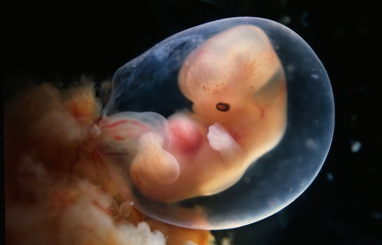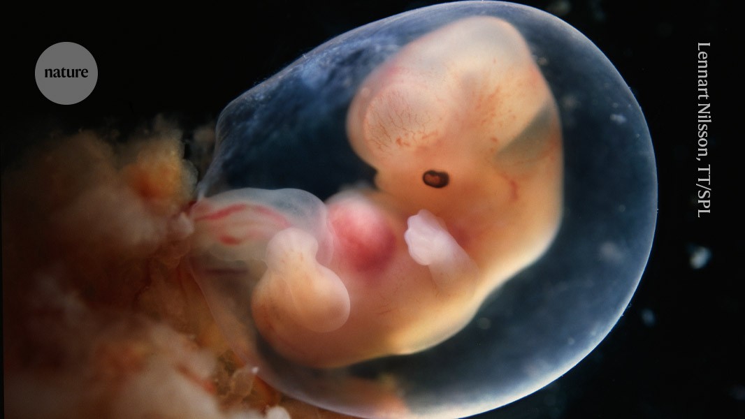
The amniotic sac protects developing embryos. Scientists have made advanced model sacs.Credit: Lennart Nilsson, TT/Science Photo Library
Researchers have coaxed stem cells to grow into amniotic sacs filled with fluid1. The model sacs, which grew to roughly the same size as a four-week-old sac surrounding a developing embryo, could be used to study the protective structure.
The amnion is a thin, transparent film that forms a fluid-filled sac that shields and cushions an embryo, potentially supporting its development. But researchers can’t easily access and study the tissue at early stages of pregnancy. Stem-cell models are a way to investigate early embryo development, but first researchers need to recreate in the laboratory what grows in the womb. The latest study, published in Cell today, is the most advanced model of the amniotic sac so far.
“The major advantage is it’s big and reproducible,” says Janet Rossant, a developmental and stem-cell biologist at the Hospital for Sick Children in Toronto, Canada, of the model. “It’s a neat paper.”

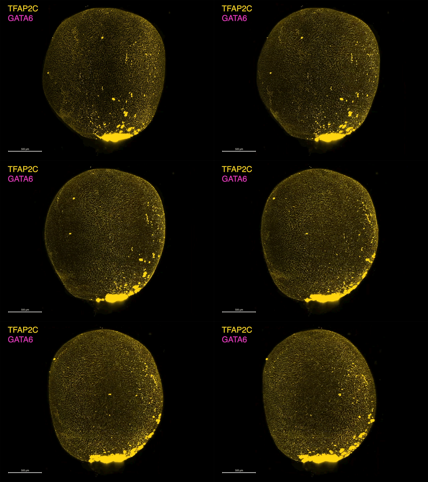
A fluid-filled amniotic sac-like structure at day 18.Credit: Gharibi et al./Cell (CC BY 4.0)
To create the model, Borzo Gharibi, a stem-cell biologist at the Francis Crick Institute in London, prodded embryonic stem cells with one type of signalling molecule for a day, and another type the next day. The cells were then placed in culture medium, and gradually formed tiny fluid-filled sacs that over three months continued to expand to roughly 2 centimeters wide. “It’s quite remarkable, the self-organization properties of these cells,” says study co-author Silvia Santos, a stem-cell biologist at the Crick.
Large sacs
On close examination, the researchers found that the cells mimicked a four-week-old amniotic sac. They contained a two-layer membrane and were filled with fluid. A structure resembling a yolk sac, which nourishes the embryo, also appeared attached to the amniotic sac, and then disappeared after two weeks.


