


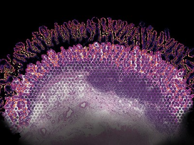
An imaging method reveals various proteins (blue, yellow and magenta) inside the nucleus of a human connective-tissue cell.Credit: Ajay Labade, Zachary Chiang, Caroline Comenho and Jason Buenrostro
Researchers are queuing up to try a powerful microscopy technique that can simultaneously sequence an individual cell’s DNA and pinpoint the location of its proteins with high resolution — all without having to crack the cell open and extract its contents. Imaging DNA and proteins inside intact cells provides crucial information about how these molecules work together.
The method’s developers have already used it to study how ageing might alter the way that proteins in the nucleus interact with chromosomes. As the body ages, they found, changes in these nuclear proteins could suppress gene activity.
“This paper is really extraordinary,” says Ankur Sharma, a cancer biologist at the Garvan Institute of Medical Research in Sydney, Australia, who was not involved in the study but is keen to use the approach to study cancer cells and described it as “phenomenal” on the social media platform, X.
The method, called expansion in situ genome sequencing, was described in a preprint1 posted on bioRxiv on 26 September. It has not yet been peer reviewed.
DNA packaging
The approach could be particularly useful to researchers who are studying how DNA is wound around proteins and stuffed into the nuclei of cells — and how the location of genes within that morass can affect their activity. We can think of DNA as “a linear string of information that has to be squished and organized inside a 5-micron-sized cell nucleus”, says Jason Buenrostro, a geneticist at Harvard University in Cambridge, Massachusetts, and an author on the preprint. “There’s a lot of information in how that folding happens.”
To extract that information, Buenrostro and his colleagues blended two previously reported methods. One feeds the cell a special enzyme for copying DNA, together with a suite of fluorescently tagged DNA components to be incorporated, one by one, into the growing DNA strands. By reading the sequence in which the fluorescent tags are added, researchers can determine the sequence of fragments of the genome2.

The new imaging technique can also be used to display genomic information. Each colour represents a different chromosome in the nucleus of the same connective-tissue cell shown above.Credit: Ajay Labade, Zachary Chiang, Caroline Comenho and Jason Buenrostro
Researchers have long known how to label proteins with tags to track their locations. But the resolution of light microscopy is limited by the wavelength of light, which makes it difficult to distinguish fluorescently tagged strands of DNA or proteins that are very close together. This poses a particular problem in the narrow confines of the nucleus.
So the team added another method called expansion microscopy3. This technique relies on a gel that permeates cells and then swells when it absorbs water — much like the filling in disposable diapers. As the gel expands, it pushes molecules further apart, making it easier to distinguish one protein molecule from one another.
The marriage of the two methods allowed Buenrostro’s team to study interactions between proteins and genes in the cells of people with Hutchinson–Gilford progeria syndrome, a genetic condition that results in premature ageing. This condition is caused by mutations in proteins called lamins, which are usually found at the periphery of cell nuclei. The researchers confirmed previous results suggesting that in individuals with progeria, these abnormal lamins intrude into the interior of the nucleus, where they seem to alter the typical arrangement of chromosomes and suppress gene activity. Similar abnormalities were present in skin cells from a 92-year-old donor who did not have progeria.
Information goldmine
Expansion in situ genome sequencing is the latest in a series of methods that are allowing researchers to collect an increasing amount of data from individual cells. The ultimate goal is to develop an approach to detect nearly any protein or metabolite in the cell, says Thierry Voet, a geneticist at KU Leuven in Belgium.
How to make spatial maps of gene activity — down to the cellular level
For now, Voet and his team are considering whether the method could be used in their studies of how cells in a developing embryo can cope with having different numbers of chromosomes from one another.
The technique requires considerable expertise, and this will limit the number of researchers who can immediately take it up, says Kelly Rogers, who studies advanced microscopy at the Walter and Eliza Hall Institute of Medical Research in Melbourne, Australia. “It definitely looks complicated.”
Even so, Rogers can list plenty of colleagues who might want to harness the approach. With time, she says, the protocols could be streamlined or even commercialized.
“One thing that is for sure is that this will become more accessible by a broader range of scientists,” says Rogers. “There don’t seem to be many limits now to what we can achieve.”