Phylum Mollusca
Punk ferox Sutton, Sigwart, Briggs, Gueriau, King, Siveter and Siveter gen. et sp. nov.
LSID. Punk: urn:lsid:zoobank.org:act:06C4B4EE-8FA2-43B6-9BB0-67DE8816C3A4. Punk ferox: urn:lsid:zoobank.org:act:2F594C68-75BB-4E62-B93E-A08BB5753FFC.
Etymology. Punk: fancied resemblance of the spicule array to the spiked hairstyles associated with the punk rock movement; ferox (Latin): wild, bold, defiant. Grammatical gender: nonbinary.
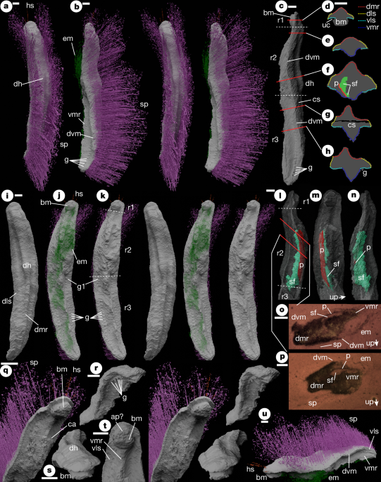
Holotype. Oxford University Museum of Natural History, OUMNH PAL-C.29644, the only known specimen (Fig. 1).
a–c,i–n,q–u, Virtual reconstructions from physical–optical dataset. d–h, Cross-sections. o,p, Optical tomograms. a, Dorsal stereo-pair. b, Left lateral stereo-pair. c, Left lateral (without spines and ?extraneous material) showing positions of regions 1–3 (white dotted lines) and cross-sections d–h (red lines). d–h, Cross-sections traced from virtual reconstruction with the extent of lateral surfaces and median ridges indicated. All scales are as shown in d. i, Dorsal stereo-pair (without spines). j, Ventral stereo-pair. k, Ventral stereo-pair (without spines and ?extraneous material) indicating regions 1–3. l, Right lateral of anterior (without spines and ?extraneous material, with body translucent) showing positions of regions 1–3 (white dotted lines) and tomograms o and p (red lines). m, Ventral of anterior (as l, scale as l). n, Left lateral of anterior (as l, scale as l). o,p, Optical tomogram Punk_B_PO_212.bmp41 (o) and Punk_B_PO_152.bmp41 (p). q, Anterior oblique stereo-pair. r, Posterior oblique stereo-pair (without spines and ?extraneous material). s, Anterior oblique (as r). t, Ventral detail of anterior (without spines and ?extraneous material). u, Left lateral oblique. ap?, putative anterior plate on buccal mass; bm, buccal mass; ca, concavity on left side; cs, concretion split; dh, dorsal ‘hump’; dls, dorsolateral surface; dmr, dorsal median ridge; dvm, dorsoventral margin; em, ?extraneous material; g, gills; g1, first (anterior-most) gill; hs, head spines; p, internal plate; r1–3, regions 1–3; sf, sediment fill; sp, spines; uc, undercut beneath buccal mass; up, younging direction indicated by sediment fill; vls, ventrolateral surface; vmr, ventral median ridge. Scale bars, 1 mm.
Stratigraphy and locality. Wenlock Series, Silurian; Herefordshire, England.
Diagnosis for genus and species. Vermiform, with dorsal and ventral median ridges. Lacking valves. Scleritome of mostly dorsally directed elongate spicules arising dorsolaterally and posteriorly. Ventral tissues non-cuticular. Approximately 25 gill pairs flanking ventral median ridge in posterior half of trunk.
Description (expanded in Supplementary Note 1). Trunk elongate and vermiform, rounded at anterior and posterior terminations (Fig. 1a–c,i–k,q–s,u). Dorsal surface with median ridge (Fig. 1i,r,s,u), broader anteriorly than posteriorly, with low ‘hump’ near midpoint (dh; Fig. 1a,i,s). Ridge flanks (concave-up in cross-section) transition laterally into dorsolateral surfaces (typically convex-up in cross-section; Fig. 1d–h); these meet at the posterior extremity of the trunk (Fig. 1r).
Valves absent. Dorsolateral surfaces bear long spines, interpreted as mineralized spicules (Fig. 1a,b,o–q,u). Spine length varies little along trunk except near terminations (Fig. 1b). Spines directed dorsally and weakly recurved towards the midline. Spine array open dorsally, except in trunk posterior (Fig. 1a). Spine array fans out anteriorly and laterally towards the trunk anterior (Fig. 1a,j,k,u). Anterior margin bears several short ‘head’ spines (Fig. 1a,j,q,u).
Dorsal surface better preserved than ventral. Surfaces separated by a sharp margin (dvm; Fig. 1c,o,p,u) differentiating dorsal integument (interpreted as cuticular) from ventral (non-cuticular). Ventral interpretation complicated by the necessarily interpretative boundary between fossil and adherent ‘extraneous material’ (rendered translucent; Fig. 1b,j,u). Ventral morphology described in three regions (r1–3; Fig. 1c,k). Region 1 is a short ‘head’ with subcylindrical ‘boss’ interpreted as buccal mass (Fig. 1c,d,j,k,q,s–u), lacking preserved opening, possibly bearing sub-semicircular anterior plate (Fig. 1t). Ventrolateral surfaces surround buccal mass and continue into region 2 (Fig. 1q,t). Regions 2 and 3 with strong median ridge between subhorizontal lateral regions (Fig. 1b,e–h,k,o–r,t,u), abutting buccal mass anteriorly, varying in cross-sectional profile along trunk. Region 2 extends to near midpoint (Fig. 1c). Asymmetrical ‘inflation’ of median ridge (Fig. 1j,k) may be a post-mortem effect. Posterior boundary of region 2 corresponds to change in width of dorsal median ridge (broader above region 2; Fig. 1c,i,k). Region 3 bears around 25 pairs of short subconical projections adjacent to median ridge, interpreted as gills (Fig. 1b,c,h,j,k,r,u).
Thin plate and geopetal cavity-fill preserved inside ventral median ridge of region 3 (Fig. 1f,l–n,o,p). Plate compositionally differentiated, probably mineralized and preserved displaced from unknown original position.
Emo vorticaudum Sutton, Sigwart, Briggs, Gueriau, King, Siveter and Siveter gen. et sp. nov.
LSID. Emo: urn:lsid:zoobank.org:act:6C11D55C-7C33-4AE4-A848-90A335C39AFF. Emo vorticaudum: urn:lsid:zoobank.org:act:8386FD34-6905-42D0-BCF6-DE196510E1EB.
Etymology. Emo: after the emo musical genre related to punk rock, whose exponents canonically bear long ‘bangs’ or fringes, of which the scleritome is reminiscent, as well as studded clothing recalling the anterior valves; vorticaudum (Latin): adjective combining vortex (whirl) and cauda (tail). The name alludes to the rotational twisting of the tail–spine arrangement. Grammatical gender: nonbinary.
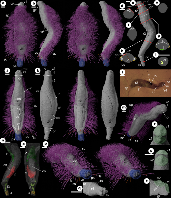
Holotype. Oxford University Museum of Natural History, OUMNH PAL-C.36023, the only known specimen (Fig. 2 and Extended Data Fig. 1).
a–c, j,k,m–t, Virtual reconstructions from synchrotron dataset. d–h, Cross-sections. l, Optical tomogram. a, Dorsal stereo-pair. b, Left lateral stereo-pair. c, Left lateral (without spines) showing positions of body divisions (white dotted lines) and cross-sections d–i (red lines). d–i, Cross-sections traced from virtual reconstruction. All scales are as shown in d (except i). j, Ventral stereo-pair. k, Dorsal stereo-pair (without spines). l, Optical tomogram Emo_A_PO_031.bmp41, oblique but sublongitudinal, positioned near part-counterpart boundary. m, Anteroventral oblique. n, Left lateral (missing anterior, without spines, with body translucent). o, Ventral (as n). p, Posterodorsal oblique stereo-pair. q, Region 4 (without spines, viewing position as left-hand p). r, Subanterior, dorsal downwards, looking directly down onto valve I (image manually enhanced to pick out valves in green, for clarity). s, Subdorsal, looking directly down onto valve II (as r). t, Lateral of anterior region (as r). br, basal ridge; cb, cuticular break (artefactual); i1–4, internal structures 1–4; gl, growth-line of valve II; gr, groove between regions 1 and 2; lp, light coloured patch in matrix; lr, lateral roll; ls, posterior lateral paired spines; mo, ?mouth; rp, respiratory protuberances/projections; ps, posterior spines; sp, main spine array; ss, ventral submedial spine; v1, valve I; v2, valve II; vr, ventral ridges; vs, postero-ventrally directed spine. Scale bars, 1 mm.
Stratigraphy and locality. Wenlock Series, Silurian; Herefordshire, England.
Diagnosis for genus and species. Vermiform, trunk lacking foot, with rounded convex dorsal and ventral surfaces. Two similar small suboval roof-like valves at anterior. Dorsal and lateral scleritome of spicular spines, absent on the head and ventrally. Articulated posterior region forming respiratory cavity surrounded by posteriorly directed spines.
Description (expanded in Supplementary Note 1). Vermiform (Fig. 2c,j,k), weakly dorsoventrally compressed (Fig. 2e–i), preserved with dorsoventral fold near midpoint (Fig. 2b,c). The body comprises four regions (r1–4 anterior to posterior; Fig. 2c,j,k).
Region 1 is a short ‘head’ bearing two similar small valves, Valve I anterior-facing, valve II dorsal-facing (Fig. 2a,k,r–t). Valves in near contact, at a high angle (Fig. 2t). Both valves suboval with distinct and weakly concave lateral areas flanking a median ridge, lacking ornament, but with one growth-line (Fig. 2t). Raised ventromedian region bearing weak elongate median hollow interpreted as the mouth (Fig. 2j,m). Region 1 separated from ventral surface of region 2 by transverse groove (Fig. 2b,t).
Region 2 is a ‘neck’ with near-parallel lateral margins (Fig. 2j,k), expanding dorsoventrally, with pronounced elongate dorsal ‘hump’ (Fig. 2c,f,m,t). Lateral, dorsal and ventral surfaces continuous (Fig. 2e,f,m). Region 3, comprising most of body-length, characterized by lateral rolls, thickenings of lateral margin separating dorsal and ventral surfaces (Fig. 2g,h,j,k,m). Lateral rolls merge anteriorly into lateral surfaces of region 2; posteriorly they meet medially on ventral surface (Fig. 2j,m). Dorsal and ventral surfaces weakly convex (Fig. 2g,h); minor transverse ridges (Fig. 2b) interpreted as in vivo deformation resulting from fold. Dorsal and ventral surfaces of region 3 lacking either preservational differentiation or sharp junction, anteriorly ventral surface continuous with that of region 2, in turn continuous with region 2 lateral and dorsal surfaces. All integument of regions 1–3 thus interpreted as cuticular; foot absent.
Regions 2 and 3 bearing array of dorsal and lateral spines interpreted as mineralized spicules (Fig. 2a,b,j,l,m,p), arising from lateral rolls (the longest spines) and the dorsal surface. Spines absent ventrally and anteriorly. Gap in posterior dorsal scleritome (Fig. 2a,p) interpreted as preservational artefact (Supplementary Note 1). Dorsally directed spines recurved medially; mid-dorsal spines ‘criss-cross’ medially (Fig. 2a,p). Four posterior spines are different in direction to nearby spines (ls, vs, ss; Fig. 2b,j,m,p; Supplementary Note 1).
Region 4 is a ‘tail’, subcircular in cross-section (Fig. 2i), tapering posteriorly from transverse basal ridge (Fig. 2c,j,k,q). The posterior surface bears poorly preserved stubby protuberances, interpreted as respiratory surfaces (Fig. 2q). Region 4 encased in posteriorly directed and rotationally twisted spine array (Fig. 2a,b,j,p) originating near basal ridge, terminating the same distance beyond respiratory projections (Fig. 2p).
Four internal structures (i1–4; Fig. 2g,n,o) are preserved. These are difficult to interpret and may, in part, be artefacts of preservation. Those in region 4 (i3, i4) may represent traces of gut and respiratory structures respectively.


