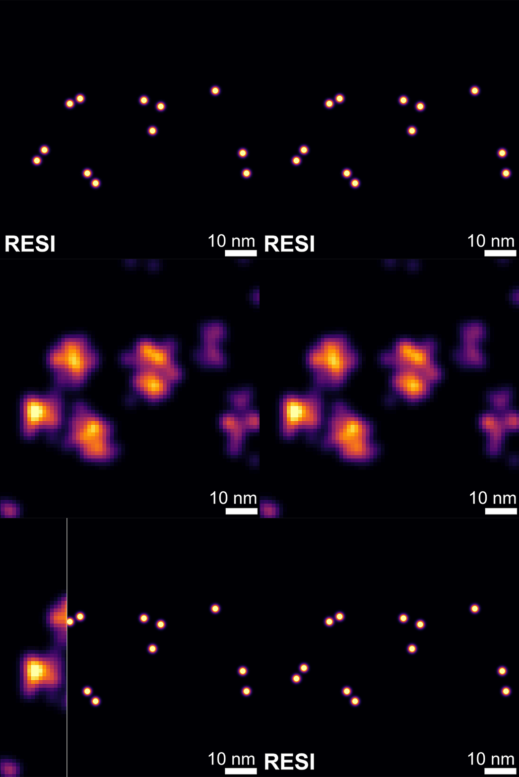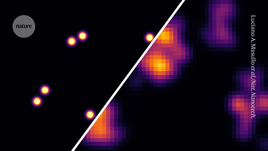
The novel 0.9-nanometre-resolution RESI imaging discriminates between molecules on a human cell membrane much more sharply than an existing 7-nanometre technique. Credit: Luciano A. Masullo et al./Nat. Nanotech. (CC BY 4.0)
Scientists have mapped individual sugar molecules on the surface of cells at a resolution once thought to be impossible for light microscopes. It is the first sub-nanometre optical resolution achieved in cells, says Sabrina Simoncelli, a chemist at University College London, who calls the results “groundbreaking”.
Every cell in the human body is wrapped in a sugary coating called the glycocalyx. It helps cells to communicate with each other and with the immune system, to fight off viruses and, in some cases, to spread cancer. Until now, no imaging tool could show the glycocalyx’s tiny sugar molecules — each of which is about one nanometre in size — in detail.
Now, researchers have found a way to image the sugars at a resolution of just 9 Ångström — 0.9 nanometres — using an off-the-shelf light microscope. In a paper published in Nature Nanotechnology on 28 July1, they reveal the arrangement of sugars on the surface of living cells that line humans’ small blood vessels.
The authors say that their imaging technique could help researchers to understand what a healthy glycocalyx looks like, and to study how it changes in diseases such as cancers and immune disorders, or in response to drugs.
“When I started my PhD, the best resolution you could get of a single sugar was a blurry image,” says study co-author Karim Almahayni, a biophysicist at the Max Planck Institute for the Science of Light in Erlangen, Germany, who completed his PhD this month. “The next step is to understand how these sugars on the cell surface change during health and disease,” he adds. “We want to judge a cell by its cover.”
Beyond the limit
Light microscopes normally blur structures smaller than 200 nanometres. Super-resolution imaging tools have achieved resolutions of 10–20 nanometres, but still lack the precision needed to distinguish between individual sugars that sit less than 10 nanometres apart in the glycocalyx.
Sugars are also notoriously difficult to tag with visible markers that make them show up under a microscope. “They cannot be genetically modified, and there are no antibodies to target them,” Simoncelli explains.
To overcome these challenges, Almahayni and his colleagues combined two Nobel prizewinning techniques. One is super-resolution fluorescence microscopy, which uses blinking fluorescent tags to map molecules; the other is click chemistry, which links two molecules and can be used inside cells.
The authors wanted to image two types of sugar — sialic acids and N-Acetyllactosamine — on the surface of cells that line blood capillaries. They started by feeding the cells modified sugars, which the cells added to their natural sugar coating. These modified sugars carried a chemical tag that allowed the researchers to attach DNA strands of six different types to the glycocalyx’s sugar molecules, which served as a docking point.


