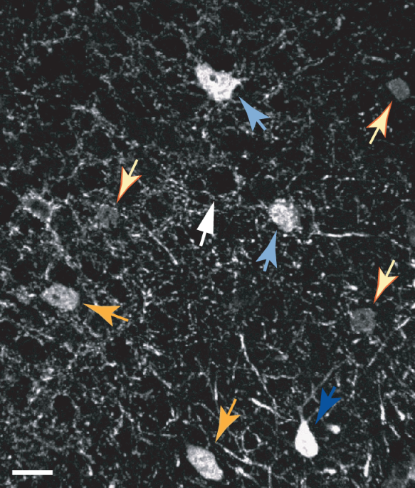
In the published version of the article, an omission occurred in Fig. 1a (left panel): the original frame around one cell highly labelled for parvalbumin (PV) was lost, which led to a failure to properly label the figure as a composite panel. This omission did not influence any of the conclusions drawn in the paper. The image was intended solely to visually illustrate the full dynamic range of PV staining intensities observable within individual experimental animals. All labelling intensity quantifications were conducted through volumetric reconstructions derived from original 3D image stacks (as explicitly detailed in the Methods section), rather than from illustrative overviews. Moreover, the analytical strategy throughout the manuscript involved pooling neuron data across multiple slices per animal within each experimental condition, as explicitly described in the main text and illustrated in Extended Data Fig. 2. This issue has been corrected in Fig. 1 below by replacing the original image with a zoomed-in view of parvalbumin immunoreactivity within the CA3 area of the hippocampus.


