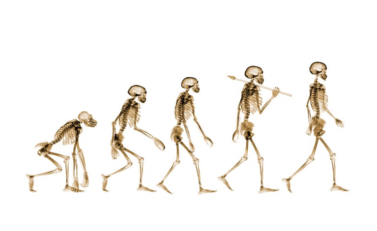
Humans have been walking on two legs for millions of years. Credit: Nick Veasey/SPL via Alamy
All vertebrate species have a pelvis, but there is only one that uses it for upright, two-legged walking. The evolution of the human pelvis, and our two-legged gait, dates back 5 million years, but the precise evolutionary process that allowed this to happen has remained a mystery.
Now, researchers have mapped the key structural changes in the pelvis that enabled early humans to first walk on two legs and accommodate giving birth to a big-brained baby. The study, published in Nature on 27 August1, compared the embryonic development of the pelvis between humans and other mammals. They found two key evolutionary steps during embryonic development — related to the growth of cartilage and bone in the pelvis — which put humans on a separate evolutionary path from other apes.
“Everything from the base of our skull to the tips of our toes has been changed in modern humans in order to facilitate bipedalism,” says Tracy Kivell, a palaeoanthropologist at the Max Planck Institute for Evolutionary Anthropology in Leipzig, Germany.
Kivell says the study offers a new understanding of how some of those changes came about, not just in living humans, but also in fossils from ancient hominins such as Denisovans. “I think it’s exciting in terms of moving forward this area of functional genomics,” she says.
Two small steps for evolution
As modern humans evolved, our pelvises developed the wide, bowl-like shape needed to allow upright, two-legged walking — but it is unclear exactly how that happened. “The human pelvis is dramatically different than what you see in chimpanzees and gorillas, so we wanted to set out to try and understand what’s happening there,” says study co-author Terence Capellini, a developmental geneticist at Harvard University in Cambridge, Massachusetts.
To investigate, the researchers studied anatomical, histological and genomic changes in samples of human pelvis from different stages of development. They then compared human pelvic development with the process in mouse embryos and other primate species, including gibbons and chimpanzees.
The researchers focussed their analysis the formation of the ilium; one of the pelvic bones that supports internal organs and anchors the gluteal muscles to stabilise walking. The team collected samples of primate embryos from museums, where they had been preserved in some cases for hundreds of years. “These museum collections are exceptionally precious; they were collected in the last hundred to two hundred years”, says Capellini.

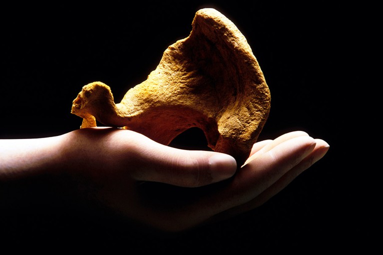
Complex genetic and molecular changes to a bone called the ilium have helped to shape the human pelvis in a way that allows us to walk on two legs.Credit: Massimo Brega/Science Photo Library
The analysis identified two key steps in the development of the human ilium which enabled its characteristic shape and therefore its ability to support bipedalism.
The first step occurs during early development of the ilium cartilage. Early bone development begins as a vertical rod of cartilage, 7 weeks after gestation. This process is similar in non-human primates. But what happens next sets the human pelvis apart from other primates — in humans, the ilium cartilage rotates 90 degrees shortly after its formation. This ultimately makes the pelvis short and broad.
The second step unique to humans occurs later in development, at 24 weeks after gestation, when the ilium cartilage ‘ossifies’ and is replaced by bone cells. In humans, some of these bone cells form much later than in other primates, which allows the cartilage cells to maintain the shape of the pelvis while it grows.
Together, these developmental quirks help to create a pelvis with the perfect shape for bipedalism.


