


Researchers used an AI model to reconstruct neural circuits in brain samples imaged using a light microscope.
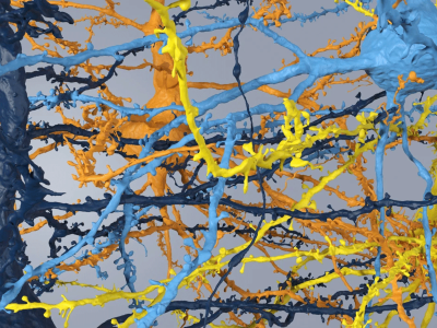
Scientists have found a way to map the intricate patterns of cells in mouse brain tissue with an off-the-shelf light microscope, using a trick that inflates a tiny sample to 16 times its original size.
Until now, charting the tangled forest of neurons in the brain — known as the connectome — required an electron microscope. These are powerful but expensive machines that cannot generate coloured images.
A milestone map of mouse-brain connectivity reveals challenging new terrain for scientists
The new approach, described in Nature on 7 May, uses gels that swell when soaked in water to space out the tightly packed neurons in a tiny piece of mouse brain, making the details visible under standard light microscopes down to individual synapses — the junctions between neurons. Using artificial intelligence (AI) models to trace individual neurons, the researchers created colourful maps that show how brain cells connect, which molecules they use to communicate and whether their signals excite or silence other cells.
“The hope is that we’re going to be able to do exactly what electron microscopy does, but faster, cheaper and in some sense more creative with the use of colour,” says Mariela Petkova, a neuroscientist at Harvard University in Cambridge, Massachusetts, who was not involved in the work.
Supersized sample
Light microscopes are cheap, fast and found in virtually all biology labs, but they lack the resolution needed to map the fine branches of neurons and synapses in the brain. The neural circuits of worms2, flies, mice and humans have all been mapped using electron microscopy.