Cytotoxin consensus sequence design
Amino acid sequences for cytotoxins were collected from the UniProt website using family: “snake three-finger toxin family Short-chain subfamily Type IA cytotoxin sub-subfamily” as a query. The resultant 86 unique CTX sequences were subjected to multiple sequence alignments in Clustal Omega62. Using these alignments, a consensus sequence was designed to represent the most common amino acid at each position across the aligned sequences. In this process, each column of the sequence alignment was analysed to select the most frequent amino acid. In scenarios in which no single amino acid was dominant, a consensus symbol was used to represent a group of similar amino acids on the basis of their properties, such as charge or hydrophobicity. This approach allowed the representation of conserved biochemical properties rather than specific amino acid identities at positions with high variability.
Secondary structure and block adjacency tensors
To generate the desired binder–target β-strand pairing interactions using RFdiffusion, fold-conditioning tensors describing single binder β-strands interacting with the target β-strands in a matrix format were supplied to RFdiffusion at inference. This information was supplied via two tensors: an [L,4] secondary one-hot tensor (0 = α-helix, 1 = β-strand, 2 = loop and 3 = masked secondary structure identity) to indicate the secondary structure classification of each residue in the binder–target complex, and an [L,L,3] adjacency one-hot tensor (0 = non-adjacent, 1 = adjacent and 2 = masked adjacency) to indicate interacting partner residues for each residue in the binder–target complex. For the design of the binders described here, the secondary structure tensor indicated an entirely masked binder structure, with the exception of binder residues set to β-strand identities, whereas the adjacency tensor indicated a masked adjacency between binder–target residues, with the exception of the predefined strand residues being adjacent to the defined target strand residues.
De novo 3FTx binder design using RFdiffusion
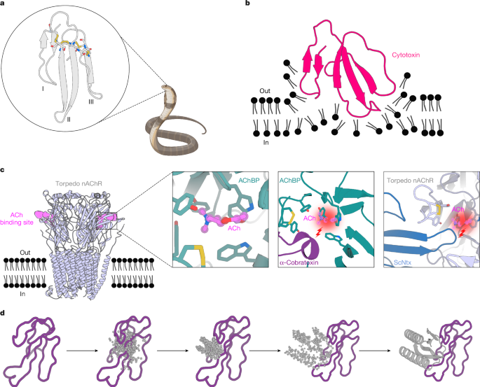
The crystal structures of ScNtx (PDB 7Z14) and α-cobratoxin (PDB 1YI5) served as the inputs for RFdiffusion. In the case of the consensus cytotoxin, the AF2 model was used. Approximately 2,000 diffused designs were generated for each target, using the secondary structure and block adjacency tensors in the RFdiffusion model. The resulting backbone libraries underwent sequence design using ProteinMPNN, followed by FastRelax and AF2 + initial guess38. The resulting libraries were filtered on the basis of AF2 predicted aligned error (PAE) < 10, predicted local distance difference test (pLDDT) > 80 and Rosetta Delta Delta G (ddg) < −40.
Partial diffusion to optimize binders
The AF2 models of the highest-affinity designs for each toxin target were used as the inputs for partial diffusion. The models were subjected to 10 and 20 noising time steps out of a total of 50 time steps in the noising schedule and subsequently denoised (“diffuser.partial_T” input values of 10 and 20). Approximately 2,000 partially diffused designs were generated for each target. The resulting library of backbones was sequence designed using ProteinMPNN after Rosetta FastRelax, followed by AF2 + initial guess38. The resulting libraries were filtered on the basis of AF2 PAE < 10, pLDDT > 80 and Rosetta ddg < −40.
Recombinant expression of ScNtx
ScNtx was recombinantly expressed from the methylotrophic yeast Komagataella phaffii (formerly known as Pichia pastoris). The ScNtx sequence was codon-optimized for expression in yeast and included an N-terminal His6 tag, followed by a biotin acceptor peptide and a tobacco etch virus proteolytic site. The expression was performed as described previously30. The culture medium was dialysed overnight against wash buffer (50 mM sodium phosphate buffer (pH 8.0) and 20 mM imidazole). Purification was carried out using an NGC chromatography system (Bio-Rad) with a 5 ml immobilized metal affinity chromatography (IMAC) nickel column (Bio-Rad). After loading, the column was washed with 5 column volumes of wash buffer to remove non-specifically bound proteins. The protein was then eluted using a gradient of 250 mM imidazole over 10 column volumes. Fractions with a high absorbance at 280 nm were pooled and dialysed against 50 mM sodium phosphate buffer (pH 8.0). Purity was assessed on SDS–PAGE to confirm the size. The protein solution was aliquoted and stored at −20 °C for further use.
Toxins
α-Cobratoxin (L8114) was obtained from Latoxan. Cytotoxin from N. pallida was obtained from Sigma-Aldrich (217503).
Venoms
Whole venoms for initial neutralization screening from N. nigricollis (CV01089563VEN) and N. pallida (CV01089566VEN) were obtained in lyophilized form from Amerigo Scientific. Catalogue numbers are provided in parentheses.
For in vitro neutralization experiments in human keratinocytes, whole venoms from N. nigricollis (L1327), Naja nigricincta (L1368), Naja mossambica (L1376), Naja nubiae (L1342), Naja katiensis (L1317), Naja ashei (L1375) and N. pallida (L1321) were purchased in lyophilized form from Latoxan. Catalogue numbers are provided in parentheses.
For the in vivo anti-cytotoxin study, N. nigricollis venom was sourced from wild-caught Tanzanian specimens housed in the herpetarium of Liverpool School of Tropical Medicine.
Gene construction of 3FTx binders
The designed protein sequences were optimized for expression in E. coli. Linear DNA fragments (eBlocks; Integrated DNA Technologies) encoding the design sequences contained overhangs suitable to cloning into the pETcon3 vector for yeast display (Addgene #45121) and LM627 vector for protein expression (Addgene #191551) through Golden Gate cloning.
Yeast display screening
For yeast transformation, 50–60 ng of pETcon3, digested with NdeI and XhoI restriction enzymes, and 100 ng of the insert (eBlocks) were transformed into Saccharomyces cerevisiae EBY100 following the protocol described in a previous study63. EBY100 cultures were cultivated in C-Trp-Ura medium with 2% (w/v) glucose. To induce expression, yeast cells initially grown in the C-Trp-Ura medium with 2% (w/v) glucose were transferred to SGCAA medium containing 0.2% (w/v) glucose and induced at 30 °C for 16–24 h. After induction, the cells were washed with PBSF (phosphate-buffered saline (PBS) with 1% (w/v) bovine serum albumin) and labelled for 40 min with biotinylated toxin targets at room temperature using the without-avidity labelling condition63. Subsequently, the cells were washed, resuspended in PBSF and individually sorted on the basis of each unique design using a 96-well compatible autosampler in the Attune NxT Flow Cytometer (Thermo Fisher Scientific).
Protein expression and purification in E.
coli for 3FTx binders
Protein expression was conducted in 50 ml of Studier autoinduction medium supplemented with kanamycin, and cultures were grown overnight at 37 °C. Cells were collected by centrifugation at 4,000×g for 10 min and resuspended in lysis buffer (100 mM Tris-HCl, 200 mM NaCl and 50 mM imidazole) supplemented with Pierce Protease Inhibitor Tablets (EDTA-free). Cell lysis was achieved by sonication using a Qsonica Q500 instrument with a four-pronged horn for 2.5 min ON total at an amplitude of 80%. Soluble fractions were clarified by centrifugation at 14,000×g for 40 min and subsequently purified by affinity chromatography using Ni-NTA resin (Qiagen) on a vacuum manifold. Washes were performed using low-salt buffer (20 mM Tris-HCl, 200 mM NaCl and 50 mM imidazole) and high-salt buffer (20 mM Tris-HCl, 1,000 mM NaCl and 50 mM imidazole) before elution with elution buffer (20 mM Tris-HCl, 200 mM NaCl and 500 mM imidazole). Eluted protein samples were filtered and injected into an autosampler-equipped ÄKTA pure system on a Superdex S75 Increase 10/300 GL column at room temperature using SEC running buffer (20 mM Tris-HCl and 100 mM NaCl (pH 8)). Monodisperse peak fractions were pooled, concentrated using spin filters (3 kDa molecular weight cutoff; Amicon; Millipore Sigma) and stored at 4 °C before downstream characterizations. Protein concentrations were determined by measuring absorbance at 280 nm using a NanoDrop spectrophotometer (Thermo Fisher Scientific) using the molecular weights and extinction coefficients obtained from their amino acid sequences using the ProtParam tool.
BLI binding experiments
BLI experiments were performed on an Octet RED96 (ForteBio) instrument, with streptavidin-coated tips (Sartorius item no. 18-5019). Buffer comprised 1× HBS-EP+ buffer (Cytiva BR100669) supplemented with 0.1% w/v bovine serum albumin. The tips were preincubated in buffer for at least 10 min before use. The tips were then sequentially incubated in biotinylated toxin target, buffer, designed binder and buffer.
Affinity measurements by SPR
SPR experiments were conducted using a Biacore 8K instrument (Cytiva) and analysed using the accompanying evaluation software. Biotinylated α-cobratoxin was immobilized on a streptavidin sensor chip (Cytiva). For ScNtx and N. pallida cytotoxin, immobilization involved the activation of carboxymethyl groups on a dextran-coated chip through reaction with N-hydroxysuccinimide. The ligands were then covalently bonded to the chip surface by means of amide linkages, and the excess activated carboxyls were blocked with ethanolamine (https://doi.org/10.1007/978-1-59745-523-7_20). Increasing concentrations of protein binders were flown over the chip in 1× HBS-EP+ buffer (Cytiva BR100669).
Circular dichroism
The secondary structure content was evaluated by CD in a Jasco J-1500 CD spectrometer coupled to a Peltier system (EXOS) for temperature control. The experiments were performed on quartz cells with an optical path of 0.1 cm, covering a wavelength range of 200–260 nm. The CD signal was reported as molar ellipticity (θ). The thermal unfolding experiments were followed by a change in the ellipticity signal at 222 nm as a function of temperature. Proteins were denatured by heating at 1 °C min−1 from 20 to 95 °C.
Crystallization and structure determination
Crystallization experiments for the binder complex were conducted using the sitting drop vapour diffusion method. Crystallization trials were set up in 200 nl drops using a 96-well format by mosquito LCP from SPT Labtech. Crystal drops were imaged using the UVEX crystal plate hotel system by JANSi. Diffraction quality crystals for the LNG binder complex appeared in 1.5 M ammonium sulfate and 25% (v/v) glycerol in 2 weeks. Diffraction quality crystals for the SHRT binder appeared in 0.08 M sodium acetate trihydrate (pH 4.6), 1.6 M ammonium sulfate and 20% (v/v) glycerol. For CYTX_B10-complex diffraction, quality crystals appeared in 0.1 M 2-(N-morpholino)ethanesulfonic acid (MES) (pH 6), 0.01 M zinc chloride, 20% (w/v) polyethylene glycol (PEG) 6000 and 10% (v/v) ethylene glycol. The crystals were flash-cooled in liquid nitrogen before being transported to the synchrotron for diffraction experiments.
The diffraction data were collected at the National Synchrotron Light Source II Beamline AMX (17-ID-1). X-ray intensities and data reduction were evaluated and integrated using XDS64 and merged/scaled using Pointless/Aimless in the CCP4i2 Program Suite65. The structure was determined by molecular replacement using a model designed using Phaser66. Following molecular replacement, the model was improved and refined using Phenix67. Model building was performed using Coot68 in between refinement cycles. The final model was evaluated using MolProbity69. Data collection and refinement statistics are reported in Extended Data Table 1. The final atomic coordinates, mmCIF and structural factors were deposited in the PDB with accession codes 9BK5, 9BK6 and 9BK7, respectively.
In vitro neutralization using electrophysiology
Human-derived rhabdomyosarcoma RD cells (American Type Culture Collection) endogenously expressing the muscle-type nAChR were used for electrophysiology experiments20. Planar whole-cell patch-clamp recordings were conducted on a Qube automated electrophysiology platform (Sophion Bioscience) with 384-channel patch chips (patch hole resistance 2.00 ± 0.02 MΩ), following the protocol detailed in a previous study20. Protein binders were preincubated with approximately one 80% inhibitory concentration (IC80) of α-cobratoxin or ScNtx at various toxin-to-binder molar ratios (1:1, 1:3, 1:9 and 1:27) and then added to the cells. The ability of the toxin to inhibit an acetylcholine (ACh; 70 µM) response in the presence or absence of binders was normalized to the full ACh response, averaged in each group (n = 16) and represented in a non-cumulative concentration–response plot. We analysed data using Sophion Analyzer v.6.6.70 (Sophion Bioscience) and GraphPad Prism v.10.1.1 (GraphPad Software).
Initial neutralization screening of whole venoms using cell viability assay
HEK293T cells were cultured in Dulbecco’s modified Eagle’s medium (Gibco) supplemented with 10% fetal bovine serum at 37 °C and 5% CO2. Cells were subjected to commercial whole venoms from N. pallida (34 µg ml−1) and N. nigricollis (42 µg ml−1), either in the absence or presence of 1:1 or 5:1 molar ratio of toxin:binder. Buffer and binder-only controls were run in parallel, and all samples were preincubated for 30 min at room temperature before addition to HEK293T cells. To determine the percentage of viable cells, RealTime-Glo MT Cell Viability Assay (Promega) was performed according to the manufacturer’s protocol. Experiments were performed in triplicate, and the results were expressed as mean ± s.d.
In vitro neutralization of whole venoms using cell viability assay
N/TERT-immortalized keratinocytes were cultured as described previously70. After determining the IC50 for seven venoms of Afronaja snakes, N/TERT cells were subjected to twice the IC50 of each venom, either in the absence or presence of a 1:5 molar ratio of venom:binder. Buffer and binder-only controls were run in parallel, and all samples were preincubated (30 min at 37 °C) before addition to N/TERT cells. To determine the percentage of viable cells, the CellTiter-Glo Luminescent Cell Viability Assay (Promega) was performed according to the manufacturer’s protocol. Experiments were performed in triplicates, and results were expressed as mean ± s.d.
LD50 determinations for α-neurotoxins
All assays used male non-Swiss albino mice (20–30 g), and all doses were mass adjusted. The toxins assayed were α-cobratoxin (7,820 Da, from N. kaouthia venom obtained from Latoxan S.A.S.) and the short-chain neurotoxin ScNtx (8,944 Da, recombinantly expressed). The toxins were solubilized in PBS at 1.0 mg ml−1 and then diluted in PBS as needed. For toxin LD50 determination, five doses with three mice per dose were used, and a 100 µl bolus was injected intraperitoneally in the right lower abdominal region; controls received only PBS. The injected mice were observed for the first 2 h and then again at 24 h. LD50 values were calculated using the Quest Graph LD50 Calculator71.
In vivo neurotoxicity protein binder protection assays
In the preincubation experiments, three LD50 values of the toxins (α-cobratoxin, 0.294 µg g−1 mouse; ScNtx, 0.261 µg g−1 mouse) were mixed with a ten-fold molar excess of their respective protein binders in PBS and incubated at room temperature for 30 min before intraperitoneal administration. Groups of five mice were injected with the binder:toxin mixture and observed at 2 and 24 h. In rescue experiments, toxins (three LD50 values) were administered intraperitoneally 15 or 30 min before the corresponding binder, given intraperitoneally at either ten-fold or five-fold molar excess to groups of five mice. Protection against lethality was measured as per cent mortality at 24 h.
In vivo dermonecrosis protein binder protection assays
CD1 male mice (18–20 g; Charles River Laboratories) were acclimated for 1 week before experimentation in specific pathogen-free conditions. The holding room conditions were 23 °C with 45–65% humidity and 12/12 h light cycles (350 lux). Mice were housed in Tecniplast GM500 cages (floor area of 501 cm2) containing 120 g LIGNOCEL wood fibre bedding (JRS) and Z-nest biodegradable paper-based material for nesting and environmental enrichment (red house, clear polycarbonate tunnel and loft). The mice had ad libitum access to irradiated PicoLab food (LabDiet) and reverse osmosis water in an automatic water system. The animals were split into cages (experimental units) upon arrival, and no further randomization was performed.
All mice were pretreated with 5 mg kg−1 morphine (injected subcutaneously) before receiving intradermal injections in a 100 μl volume into the ventral abdominal region (rear side flank region). A venom-only control group of five mice received 63 μg of N. nigricollis (Tanzania) venom (dissolved in PBS). For protection assays, crude venom was preincubated (30 min at 37 °C) with varying cytotoxin:binder ratios of 1:1, 1:2.5 and 1:5 before injection (n = 3) (ratios estimated from the proportion of cytotoxin in the venom). Before this, the control group (N = 3) received injections of cytotoxin binder alone (278 µM, equivalent to the 1:5 cytotoxin:binder dose) to check tolerance of the cytotoxin binder. For sample size, N = 3 was used for groups receiving the cytotoxin binder because this was a pilot experiment. N = 5 was used for the venom-only control group because of the variation in lesion size, which is the size recommended by the World Health Organization. In total, 17 mice were used. No inclusion or exclusion criteria were used during the experiment, and all data points were used in the analysis. No strategy was used to control confounders. All experimenters were aware of the group allocation during the experiment and analysis.
After 72 h, the mice were euthanized with increasing concentrations of CO2, and the lesions were excised. The outcome measured was the lesion size. Photographs of the lesions were taken using a digital camera immediately after excision, and the severity and size of the dermonecrotic lesions were determined using Venom Induced Dermonecrosis Analysis tooL (VIDAL)72.
Ethical approval
Animal experiments for in vivo neurotoxicity assays were conducted at the University of Northern Colorado under protocol 2303D-SM-S-26, approved by the University of Northern Colorado Institutional Animal Care and Use Committee (UNC-IACUC), in accordance with Government Principles, Public Health Policy, US Department of Agriculture Animal Welfare Act and the Guide for the Care and Use of Laboratory Animals.
Animal experiments for in vivo dermonecrosis assays were approved by the Animal Welfare and Ethics Review Board of the Liverpool School of Tropical Medicine and the University of Liverpool, and conducted under the UK Home Office project licence P58464F90 in accordance with the UK Animal (Scientific Procedures) Act 1986.
Cell line development, acquisition and authentication
HEK293T cells (American Type Culture Collection CRL-3216) and N/TERT-immortalized keratinocytes, provided by E. O’Toole (Queen Mary University of London), were authenticated by means of morphological assessment and tested for mycoplasma contamination.
Reporting summary
Further information on research design is available in the Nature Portfolio Reporting Summary linked to this article.


