


Under President Donald Trump, the US government is gutting scientific research. The National Institutes of Health has cut almost US$2 billion of grants that were already approved, and the National Science Foundation has terminated more than 1,400 grants. And the president has even bigger plans to eviscerate science. His proposed budget for fiscal year 2026 would cut non-defence-related research and development by 36%.
“They have cancelled wholesale a wide variety of research efforts in midstream,” says John Holdren at Harvard University in Cambridge, Massachusetts, who was the science adviser to former president Barack Obama during both his terms. “They intend to now solidify it with cuts in the budgets.”
The cancelled and threatened research is a mixture of ‘applied’ work that has stated applications, which can be commercial in nature, and ‘basic’ or ‘blue skies’ research, intended to develop new knowledge.
From MRI to Ozempic: breakthroughs that show why fundamental research must be protected
Basic research is easily mocked because it can seem impractical, but, in fact, it is a major driver of economic growth. “The return on investment in basic research — the return to society — is very high, typically multiple dollars back per dollar invested,” says Holdren.
The US funding cuts will hit basic research particularly hard because the government has historically been the prime supporter of fundamental research. The private sector will never invest enough in such research, says Holdren. “The timescale for returns is too long and the ability of the funder to capture those returns too uncertain,” he says. “That’s the reason that the funding of fundamental research is, at its base, a responsibility of government.”
Although it’s impossible to estimate how the reductions in federal support might limit future discoveries, scientists point to a long list of findings that emerged out of fundamental research and went on to change the world. Here are a few examples.
From hot springs to DNA forensics
In the summer of 1966, while he was an undergraduate at Indiana University, Hudson Freeze went to live in a cabin on the edge of Yellowstone National Park. He was working for microbiologist Thomas Brock, who was convinced that certain microorganisms were living at surprisingly high temperatures. Dodging bears, and the traffic jams they caused, Freeze visited the hot springs every day to sample their bacteria.

Thomas Brock stands by Mushroom Spring in Yellowstone National Park in 1967. Bacteria that thrive in high-temperature fluids were discovered at the site.Credit: Thomas Brock/USGS
On 19 September, Freeze succeeded in growing a sample of yellowish microbes from Mushroom Spring. Under a microscope, he found an array of cells collected from the near-boiling fluids. “I was seeing something that nobody had ever seen before,” says Freeze, now at the Sanford Burnham Prebys Medical Discovery Institute in La Jolla, California. “I still get goosebumps when I remember looking into the microscope.”
Three years later, Freeze and Brock described one of the bacteria, which they called Thermus aquaticus1. It grew best at 70 °C. Then, in 1970, they isolated an enzyme2 from T. aquaticus, which performed sugar metabolism at an optimal temperature of 95 °C. By then, Freeze had left for graduate studies and shifted his priorities to slime moulds. However, other researchers kept studying T. aquaticus, and in 1976, a team at the University of Cincinnati in Ohio isolated another enzyme3: a ‘DNA polymerase’ that could synthesize new DNA at 80 °C.

How Trump 2.0 is reshaping science
Seven years later, this Taq polymerase would prove to be just what biochemist Kary Mullis needed to create the polymerase chain reaction (PCR), a method for rapidly making thousands upon thousands of copies of a single fragment of DNA4. Mullis needed high temperatures to break DNA molecules apart, so he also needed a polymerase that worked at high temperatures to avoid repetitive heating and cooling.
Today, PCR is an indispensable tool in everything from medicine — where it’s used to match organ donors with recipients and in the diagnosis of cancers — to DNA fingerprinting that can help police to identify killers.
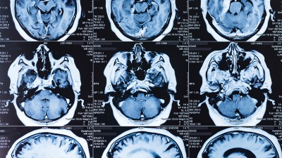
The origins of MRI
Magnetic resonance imaging (MRI) is a mainstay of modern medicine. It can generate detailed images of a person’s internal anatomy — revealing, for instance, abnormal structures in the heart or whether a tumour has grown or shrunk. A variant called functional MRI (fMRI) tracks changing blood flow in the brain, which has enabled researchers to discover fundamental insights about how the brain works. What’s more, MRI is non-invasive and doesn’t require the use of radioactive substances or ionizing radiation, unlike many other imaging methods.
MRI emerged from research in the 1930s into the physical properties of atomic nuclei, and of the fundamental particles within them. It was “pretty esoteric stuff” with “no applications really in sight or in mind”, says Carmen Giunta, a chemist at Le Moyne College in Syracuse, New York.

Research in the 1930s by Isador Rabi eventually helped in the development of MRI scanning technology.Credit: RDB/ullstein bild/Getty
One key discovery on the path to MRI machines involved studies of protons and neutrons, which make up atomic nuclei. Such particles have a property called spin that describes their angular momentum.

Will US science survive Trump 2.0?
In the 1930s, physicist Isidor Rabi and his colleagues were investigating spin by firing beams of atomic nuclei through magnetic fields. Protons and neutrons, depending on the orientations of their spins, have slightly different energy levels when exposed to magnetic fields. “The resonance method that he developed was a way of detecting when these spins change their orientation in the presence of a magnetic field,” says Giunta. For this work, Rabi won the Nobel Prize in Physics in 1944.
Nuclear magnetic resonance found its first uses in chemical laboratories. Because atomic nuclei are sensitive to their surroundings, careful measurements of magnetic resonance could be used to determine how atoms were connected in large molecules. From the 1970s onwards, it was fashioned into a tool for imaging biological tissues. Paul Lauterbur and Peter Mansfield shared the Nobel Prize in Physiology or Medicine in 2003 for their work in developing MRI.
Root vegetables and flat-screen TVs
It started in Prague in early 1888, when botanist Friedrich Reinitzer extracted chemicals called cholesterol esters from carrot roots. One of the substances — crystals of cholesteryl benzoate — did something unexpected. Normal crystals, when heated, lose both their solidity and colour at the same temperature, but these did not. “They lost their solidity at 145 °C but retained their bluish colour, which they only lost at 178 °C,” says Michel Mitov at the University of the Côte d’Azur in Nice, France.
Other researchers had seen similar behaviours before, but Reinitzer recognized that there might be an important new phenomenon at work. Unsure how to explain it, on 14 March he wrote a long letter to physicist Otto Lehmann in Aachen, in what is now Germany. “Lehmann was the perfect colleague for continuing and reproducing the observation,” says Mitov, because he had built a microscope with a heated stage, which meant he could observe the crystals’ behaviour in real time. The two exchanged letters and samples for weeks, and Reinitzer presented their initial results at a meeting in Vienna in May.

Otto Lehmann made key discoveries about liquid crystals, which helped to lay the foundation for modern screens on televisions and smartphones.Credit: Unknown author
Lehmann’s key observation was that, when the crystals lost their solidity, they still retained some properties of a crystal. Yet in other respects they were liquid. At a molecular level, they were composed of long molecules that remained in an ordered orientation (as in a crystal) but could also move freely (as in a liquid). Lehmann called them liquid crystals.
For decades, many researchers refused to accept this, because it flew in the face of the system that physicists and chemists used to categorize matter. Substances were either solid, liquid or gaseous. Liquid crystals blurred the lines, and acceptance of this came at “a very high intellectual cost”, says Mitov.
In the first half of the twentieth century the evidence became undeniable, but research into liquid crystals dried up, out of a belief that they would never be of any use. The field was revived by US chemists in the late 1950s, and in 1968, engineers developed the first flat screens based on liquid crystals, which ultimately gave rise to flat-screen televisions. However, their applications go far beyond screens, says Mitov, including uses in cameras, microscopes, smart materials, robotics and even anti-counterfeiting technology.
The tiny start to gene editing
“My head explodes every time I see a new application of CRISPR, or that CRISPR has cured someone,” says microbiologist Francisco Mojica at the University of Alicante in Spain.

US Supreme Court allows NIH to cut $2 billion in research grant
CRISPR, short for Clustered Regularly Interspaced Short Palindromic Repeats, is a tool that can edit genomes in a very precise way. It opened up vast opportunities for basic research and has paved the way for curing genetic disorders, including sickle-cell disease, immune dysfunction and life-threatening metabolic conditions. Emmanuelle Charpentier and Jennifer Doudna shared the 2020 Nobel Prize in Chemistry for developing the tool.
The discoveries that led to this revolution happened decades earlier. In 1989, Mojica was a PhD student investigating Haloferax mediterranei R-4: a single-celled organism called an archaean, found in salt-producing ponds near Alicante. He tried to find out how the microbe could survive in such briny conditions. Having identified some promising regions of the microbe’s genome, Mojica sequenced them and was surprised to find short segments that were repeated at regular intervals. He and other researchers gave these repeated segments different names but eventually settled on the acronym CRISPR. As he investigated them, Mojica proposed some potential functions for the repeating segments: ideas that were, he says wryly, “absolutely wrong”5.

Francisco Mojica’s studies of microbial genes underpinned the development of the CRISPR gene-editing system.Credit: Juan Carlos Soler/ARCHDC/Archivo ABC/Alamy
Subsequently, Mojica learned that similar sequences could be found in many other microbes, which didn’t live in saline conditions. “Whatever role they were playing, it couldn’t be related to the peculiarities of the diverse environments,” he says.
The key clue was the discovery, between the repeated segments, of sequences from the genomes of bacteriophages: viruses that infect bacteria. Eventually Mojica realized that a bacterium carrying a sequence from a specific phage could not be infected by that phage. “We inferred that this was an adaptive immune system,” he says. “One ancestor acquired spacers from the phage, and after that, the descendants were resistant to infection6.”
Mojica knew this was a big find — an adaptive immune system had never been observed in bacteria or archaea. He also thought that it might be useful for dealing with bacterial infections. Then others found that CRISPR works by cutting DNA at specific points7. From there, Doudna and Charpentier discovered how they could harness that system and reprogram it to edit genes. And so the CRISPR revolution took off8.
Weight loss, inspired by a lizard
Weight-loss and diabetes medications such as Ozempic have become the wonder drugs of the era. Almost 5% of people in the United States have used them to slim down, and the global market for these drugs is expected to reach $100 billion by 2030. Although much of the work that led to them was done for medical applications, one key discovery came from research on the only venomous lizards in the United States: Gila monsters (Heloderma suspectum).

A Gila-monster peptide played a part in the development of GLP-1 drugs.Credit: Getty
At the heart of the story is a molecule called glucagon-like peptide-1 (GLP-1), which is produced in the human gut. In the 1980s, chemist Svetlana Mojsov showed that GLP-1 can stimulate the production of insulin, lowering blood sugar levels9.
Daniel Drucker, now at the University of Toronto in Canada, worked with Mojsov on this early research. “We were focused on its potential for diabetes treatment,” he says. “Ten years later, in 1996, we and others discovered that GLP-1 reduces food intake, and the potential for weight loss emerged.” However, there was a problem. GLP-1 has a short half-life: just a few minutes. That meant it was no use as a drug, because it would be broken down in the body before it could have much of an effect.